Download
1 / 77
770 likes | 866 Views
Learn the management of stages of labor, pain relief, and fetal assessment at King Khalid University Hospital's Obstetrics & Gynecology Department. Understand labor definition, cervical dilatation, effacement, and stages of labor. Dive into maternal and fetal systems during labor, the second stage mechanics, crowning, and episiotomy types. Gain insights into the third stage and placental delivery. This course covers all aspects of labor management and fetal assessment, preparing you comprehensively. Practice your knowledge for better maternal and fetal outcomes.

E N D
Management of labour & fetal assessment King Khalid University Hospital Department of Obstetrics & Gynecology Course 482
Management of labour & fetal assessment Objectives: Managements of the stages of labour Pain relief in labour Fetal assessment (antenatal & intra-partum)
Management of labour Definition of labour: Progressive cervical effacement and dilatation resulting from regular uterine contractions that occur at least every 5 minutes and last 30-60 seconds Braxton Hicks: contractions Not associated with cervical changes Lightening: Descent of the fetal head into the pelvis There are 4 stages of labour
First stage of labour Start from onset of true labour pain----full dilatation of cervix In primigravida------ 12 hour duration In multigravida-----6 hours duration Chiefly concerned with preparation of the birth canal as to facilitate expulsion of the fetus in the second stage It has 2 phases A latent phase up to 3 cm dilatation of cervix • is variable: up to 8 hours in primi • 4 hours in multi An active phase from 3 cm to full dilatation of cervix Rate of dilatation 1 cm/hour in primigravida • 1.5 cm/ hour in multigravida
Dilatation of the cervix Dilatation usually measured by fingers but recorded in cm Dilatation relates with dilatation of internal os
Effacement or taking up of cervix Muscle fibers of cervix are pulled upward and merges with the fibers of the lower uterine segment Cervix becomes thin during first stage In primi----- effacement precedes dilatation of the cervix In multi-----both occur simultaneously Effacement is determined by the length of the cervical canal in the vagina Effacement is expressed in terms of percentage
First stage of labour Maternal system Fetal system -As so long as the membranes are intact, usually there is no adverse effect on the fetusBUT However, during contraction there may be slowing of FHR by 10-20 bpm which soon returns to its normal as the intensity of contraction diminishes -General condition remains unaffected -Pulse rate increases by 10-15 bpm during contraction with the settle down to its previous rate in between contractions -Systolic BP increase by 10 mm Hg during contraction - Temperature remains unaffected
Management of labour Initial assessment: History: Onset, strength, frequency of contractions Leakage of fluid Vaginal bleeding Fetal movement Medications Last oral intake Review of past obstetric history, prenatal lab tests, gestational age, parity, size of previous infants, any antenatal complications
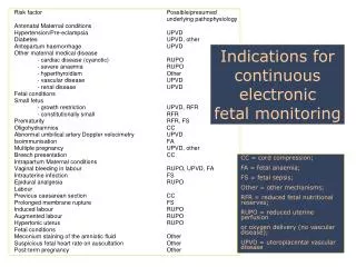
Management of the first stage of labour • -Informed consent on management of labour & delivery • Maternal position---lateral recumbent position • Avoid supine hypotension • Partogram: -Iv fluids & avoid oral intake -Maternal vital signs every 1-2 hours -Input-output monitoring -Analgesia -Fetal heart rate monitoring (CTG) -Uterine contractions monitoring -Vaginal examination for cervical dilatation & poistion in active phase every 2 hours • -Amniotic membranes status & amniotic fluid colour
Mechanics of labour The Power: force generated by uterine contraction
Second Stage of labour From full dilatation of cervix till delivery of the neonate The mother has a desire to bear down with each contraction Last from 30 minutes to 3 hours in primigravida 5-30 minutes in multigravida
Management of the second stage of labour Molding (alteration of the relationship of the fetal cranial bones to each other as a result of compression forces by the bony pelvis) Caput (localized edematous swelling of the scalp caused by pressure of the cervix on the presenting portion of the fetal head)--- gives false impression of fetal descent
Management of the second stage of labour Crowning ( when the largest diameter of the fetal head is encircled by the vulvar ring) -Vaginal examination every 30 minutes -Maternal position– any comfortable position for bearing down -Bearing down---with each contraction -Delivery of the fetal head---manual perineal support -Fetal airway clearance -Umbilical cord clamping -Place the infant under warmer
Episiotomy Incision in the perineum after crowning to aid delivery and avoid laceration of perinium Types: Right mediolateral Left mediolateral Central
PERINEAL LACERATION 4 TYPES: -First degree: laceration involving the vaginal epithelium or perineal skin -Second degree: laceration extending into the sub-epithelial tissues of the vagina or perineum with or without involving the perineal body Third degree: laceration involving anal sphincter Fourth degree: laceration involving rectal mucosa
Third stage of labour The interval between the delivery of the infant and complete delivery of the placenta & membranes Duration is 5-30 minutes Signs of placental separation: 1- Fresh blood show from vagina 2- The umbilical cord lengthens outside the vagina 3- The fundus of the uterus rises up 4- The uterus becomes firm & globular The placenta should be examined to ensure that it is complete The blood loss should be estimated
Forth stage of labour The hour immediately after the delivery • Needs close observation of: blood pressure, pulse rate, uterine blood loss Watch for post partum hemorrhage
Pain relief in labour Goal: effective pain relief to the mother that is safe for her & the fetus with minimal side effects on the progress & outcome of labour
Pain relief in labour Non pharmacological method: Back massage Acupuncture Hypnosis Breathing exercises
Pain relief in labour Pharmacological methods: Narcotic analgesics– cross the placenta – cause fetal respiratory depression (Nitrous oxide, pethidine) Epidural analgesia: The most effective Contra indicated if-coagulo-pathy, infection at needle site, severe hypo-volemia Side effects: Hypotension, headache, impaired ability to push, prolonged second stage (15 Minutes) Pudendal block: for S2-S4 for the second stage of labour for instrumental delivery
Fetal assessment Aim: Ensure fetal wellbeing ( Identify patients at risk of fetal asphyxia) To prevent prenatal mortality & morbidity
Screening for high risk pregnancy History * Age *Social burden *Smoking *Past medical conditions e.g D.M, HTN *Past Obstetric history
FETAL AND NEONATAL COMPLICATIONS OF ANTEPARTUM ASPHYXIA Stillbirth (Mortality) Metabolic acidosis at birth Hypoxic renal damage Necrotizing enterocolitis Intracranial haemorrhage Seizures Cerebral palsy
CONDITIONS ASSOCIATED WITH INCREASEDPERINATAL MORBIDITY/MORTALITY Small for gestational age fetus Decreased fetal movement Postdates pregnancy (>294 days) Pre-eclampsia/chronic hypertension Pre-pregnancy diabetes Insulin requiring gestational diabetes Preterm premature rupture of membranes Chronic (stable) abruption
When to start fetal Assessment antenatally ** Risk assessed individually **For D.M. fetal assessment should start from 32 weeks onward if uncomplicated ***If complicated D.M. start at 24 weeks onward **For Post date pregnancy start at 40 weeks **For any patient with decrease fetal movement start immediately ** Fetal assessment is done once or twice weekly
Antenatal Fetal Assessment Fetal movement counting Non stress test Contraction stress test Ultrasound fetal assessment Umbilical Doppler Velocimetry
Fetal movement counting Cardiff technique: *Done in the morning, patient should : calculate how long it takes to have 10 fetal movement **10 movements should be appreciated in 12 hours
Fetal movement counting Sadovsky technique: -For one hour after meal the woman should lie down and concentrate on fetal movement -4 movement should be felt in one hour -If not , she should count for another hour -If after 2 hours four movements are not felt, she should have fetal monitoring
Non stress test *Done using the cardiotocometry with the patient in left lateral position **Record for 20 minutes
Non stress test *The base line 120-160 beats/minute *Reactive: At least two accelerations from base line of 15 bpm for at least 15 sec within 20 minutes Non reactive: No acceleration after 20 minutes- proceed for another 20 minutes
Non stress test If non reactive in 40 minutes---proceed for contraction stress test or biophysical profile The positive predictive value of NST to predict fetal acidosis at birth is 44%
Contraction stress test Fetal response to induced stress of uterine contraction and relative placental insufficiency Should not be used in patients at risk of preterm labor or placenta previa Should be proceeded by NST
Contraction stress test Contraction is initiated by nipple stimulation or by oxytocin I.V. The objective is 3 contractions in 10 minutes If late deceleration occur-----positive CST
Interpretation of CTG Normal Baseline FHR 110–160 bpm – Moderate bradycardia 100–109 bpm – Moderate tachycardia 161–180 bpm – Abnormal bradycardia < 100 bpm – Abnormal tachycardia > 180 bpm
Deceleration EARLY : Head compression LATE : U-P Insufficiency VARIABLE : Cord compression Primary CNS dysfunction
TachycardiaHypoxiaChorioamnionitisMaternal fever B-Mimetic drugsFetal anaemia,sepsis,htfailure,arrhythmias
Ultrasound fetal assessment Assessment of growth Biophysical profile (BPP)
Assessment of fetal growth by ultrasound Biometry: Biparietal diameter (BPD) Abdominal Circumference (AC) Femur Length (FL) Head Circumference (HC) Amniotic fluid Placental localization
BPD Assessment of fetal growth by ultrasound AC FL