Cystitis & Pyelonephritis
Cystitis & Pyelonephritis. Dr. Abdelaty Shawky Assistant professor of pathology. * Predisposing factors: . B ladder calculi. U rinary obstruction. D iabetes mellitus. Instrumentation. immune deficiency. Patients receiving cytotoxic antitumor drugs , such as cyclophosphamide.

Cystitis & Pyelonephritis
E N D
Presentation Transcript
Cystitis & Pyelonephritis Dr. Abdelaty Shawky Assistant professor of pathology
* Predisposing factors: • Bladder calculi. • Urinary obstruction. • Diabetes mellitus. • Instrumentation. • immune deficiency. • Patients receiving cytotoxic antitumor drugs, such as cyclophosphamide. • Women are more likely to develop cystitis as a result of their shorter urethra.
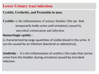
* Causative organisms: • The common etiologic agents of cystitis are: Escherichia coli, Proteus, Klebsiella, and Enterobacter. • Tuberculouscystitis is almost always a sequel to renal tuberculosis.
Candida albicans (Monilia) and, much less often, cryptococcal agents cause cystitis, particularly in immunosuppressed patients or those receiving long-term antibiotics. • Schistosomiasis (Schistosomahaematobium) is common in certain Middle Eastern countries, notably Egypt. • Viruses (e.g., adenovirus), Chlamydia, and Mycoplasma may also be causes of cystitis.
* Clinical presentation of cystitis: - All forms of cystitis are characterized by a triad of symptoms: 1. Frequency, which in acute cases may necessitate urination every 15 to 20 minutes. 2. Lower abdominal pain localized over the bladder region or in the suprapubicregion. 3. Dysuria: pain or burning on urination.
Systemic signs of inflammation such as elevation of temperature, chills, and general malaise.
Cystitis may be preceded by pyelonephritis. • Cystitis is sometimes a secondary complication of some underlying disorder such as prostatic enlargement, cystocele of the bladder, calculi, or tumors. These primary diseases must be corrected before the cystitis can be relieved.
* Types of cystitis: • Hemorrgaic cystitis. • Suppurative cystitis. • Follicular cystitis. • Eosinophilic cystitis. • Interstitial cystitis (Hunner’s ulcer). • Polypoid cystitis. • Chronic cystitis.
Hemorrhagic cystitis: • This form of cystitis follows radiation injury or antitumor chemotherapy (cyclophosphamide). • Adenovirus infection also causes a hemorrhagic cystitis. • Characterized by marked hemorrhage and ulcerations.
2. Suppurative cystitis: • Caused by pyogenic bacterial infection. • Accumulation of large amounts of suppurative exudate . 3. Follicular cystitis: Characterized by the aggregation of lymphocytes into lymphoid follicles within the bladder mucosa and underlying wall.
4. Eosinophilic cystitis: • Manifested by infiltration of the submucosa by eosinophilstogether with fibrosis and occasionally giant cells. • Most cases of eosinophilic cystitis represent nonspecific subacute inflammation, although, rarely, these lesions are manifestations of a systemic allergic disorder.
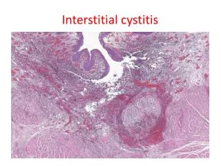
5. Interstitial Cystitis (Hunner Ulcer): • This is a persistent, painful form of chronic cystitis occurring most frequently in women and associated with inflammation and fibrosis of all layers of the bladder wall. • The condition is of unknown etiology but is thought to be of autoimmune origin, particularly because it is sometimes associated with other autoimmune disorders.
It is characterized clinically by intermittent, severe, suprapubic pain, frequency, urgency, hematuria, and dysuria without evidence of bacterial infection. • Some but not all patients exhibit morphologic features of chronic mucosal ulcers (Hunner ulcers).
* Microscopically: Inflammatory cells and granulation tissue involve the mucosa, lamina propria, and muscularis mucosa, with prominent mast cells.