Download

1 / 115
1.16k likes | 1.26k Views
Learn about the history, classification, diagnosis, and management of desquamative gingivitis, including its clinical significance and diseases mimicking its presentation. Explore the systematic approach to diagnosing this condition through clinical history, intraoral examination, biopsy, microscopic examination, immunofluorescence, and more.

E N D
Contents • History • Classification • Diagnosis of desquamative gingivitis: a systematic approach • Clinical History • Intraoral Clinical Examination • Assessment of Extraoral Involvement • Biopsy. • Microscopic Examination • Immunofluorescence • Management.
Diseases clinically presenting as desquamative gingivitis • Lichen planus • Pemphigoid • Pemphigus vulgaris • Chronic ulcerative stomatitis • Linear IgA disease (linear iga dermatosis) • Dermatitis herpetiformis • Lupus erythematosus • Erythema multiforme • Drug eruptions • Miscellaneous conditions mimicking desquamative gingivitis • Clinical significance and implications
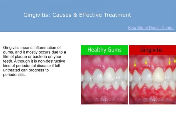
DESQUAMATIVE GINGIVITIS • Chronic desquamative gingivitis is a clinical entity characterized by erythematous and desquamative involvement of the free and attached gingiva
Prinz, in 1932, described the condition in detail and gave it the name "chronic diffuse desquamative gingivitis." • This condition was considered to be rare • chronic diffuse inflammation of the marginal gingiva characterized by desquamation of the epithelium of the papillae and adjacent gingiva. • The exposed connective tissue had a deep bluish-red color and bled upon the slightest irritation
Classification of the Periodontal Diseases American Academy of Periodontology (AAP) • A. Mucocutaneous lesions 1. Lichen planus 2. Pemphigoid 3. Pemphigus vulgaris 5. Lupus erythematosus 6. Drug induced 7. Other
DIAGNOSIS OF DESQUAMATIVE GINGIVITIS: A SYSTEMATIC APPROACH Clinical History • The onset and progression of gingival lesion • The presence of prodromal signs and symptoms • Infections and drug intake • Topically applied substances • Patient complaints • A standardized manner for collecting symptoms • The medical history • The evaluation of systemic conditions
Intraoral Clinical Examination • The extent and degree of gingival involvement vary. It can be staged. • Mild form • Moderate form • Severe form • Oral Lichen Planus lesions • Mucous Membrane Pemphigoid and Pemphigus Vulgaris • Recognition of the pattern of distribution of the lesions
Type of bulla (flaccid or tense), • Rapid bursting of the bullae • Reaction to pressure • Appearance (cicatricial pemphigoid and lichen planus tend to be more hemorrhagic; pemphigus vulgaris ulcers are shallow). • The patient with a positive nikolsky sign may facilitate the diagnosis of pemphigus vulgaris.
Identify dental materials and their topographic relationship with lesions. • Many dental restorative materials (amalgam, composites, cobalt, and gold) were reported to induce oral lichenoid reactions. • They resemble OLP clinically, histologically, and with immunofluorescence staining but may resolve after material replacement. • Such reactions should be suspected, especially when lesions are confined to small areas and are in close contact with dental materials.
Assessment of Extraoral Involvement • Skin, scalp, nails, and mucosae with squamous differentiated epithelium, such as laryngeal, esophageal, nasal, genital,and conjunctival, represent possible locations • The skin and the other mucosae should be evaluated by specialists to determine the presence, morphology, and distribution of lesions. • Some DG-associated disorders may have internal organ involvement (LE, EM, and GVHD) or may be associated with systemic disease (DH). Therefore, any systemic sign/symptom should receive attention and should be investigated adequately .
Biopsy • Given the extent and number of lesions that may be present in a given individual, an incisional biopsy is the best alternative to begin the microscopic and immunologic evaluation.
Microscopic Examination. • The epithelium may lack keratinization: • The epithelial-connective tissue interface may appear as a straight line • The epithelium may be edematous with widened intercellular spaces. • Basal cells may show lytic destruction. • Inflammatory cells of all types are seen in the lamina propria and in the epithelium before the bullae form. • The region of the basement membrane may be altered because of presence of anti bodies to the bm.
Collagen fibers may be slightly separated. • There is decreased number of anchoring fibrils. • The basal lamina may be replicated. • The subepithelial connective tissue may be vacuolated and disorganized. • The basal lamina in the region of a bulla has been reported to be adherent to the basal cells, the connective tissue, or both • Pseudopodia from the basal cells enter the bullae. • The bullae may contain fibrin, red blood cells, and cellular debris.
Management • Once the diagnosis is established, the dentist must choose the optimum management for the patient. • This is accomplished according to three factors: (1) practitioner's experience, (2) systemic impact of the disease, and (3) systemic complications of the medications. • A detailed consideration of these three factors dictates three different scenarios.
When oral treatment is provided, periodic evaluation is needed to monitor the response of the patient to selected therapy. • Initially, the patient should be evaluated at 2-4 weeks after beginning treatment to ensure that the condition is under control. This observation should continue until the patient is free of discomfort. • Appointments every 3 to 6 months would then be appropriate, of medication(s) are usually adjusted during this interval.
COMMON THERAPIES • Antimicrobials • Tetracycline usually in conjunction with nicotinamide • Nicotinamide inhibits polymorphonuclear leucocytes ,eosinophil chemotaxis and blocking mast cell degranulation byIgE mediated reactions. • Besides its antimicrobial activity, tetracycline stabilizes connective tissue.
Azathioprine • Immunosuppressive agent. It is a synthetic purine analogue which inhibits both RNA and DNA purine biosynthesis and thus inhibits cell division.. • It also has anti inflammatory properties with inhibition of T cell function and to a lesser extent B cell function. • often utilized in conjunction with other drugs for treatment of diseases including bullous pemphigoid, erythema multiforme, lupus erythematosus, pemphigus, and psoriasis.
Cyclophosphamide • cytotoxic and immunosuppressive agent that is administered either orally or intravenously. • It suppresses B cells more than T cells with T suppressor cells affected more than T helper cells. • Because of the risks of serious adverse side effects, its use is usually reserved for more serious disorders such as pemphigus.
Cyclosporine • A fungal-derived cyclic polypeptide that has been used for prevention of transplant rejection, treatment of T cell disorders, and treatment of autoimmune diseases • Its major effect is the immuno-suppression of T helper cells • Cyclosporine has been utilized for treatment of diseases including epidermolysis bullosa. acquisita, lichen planus, lupus erythematosus, pemphigus vulgaris, and psoriasis.
Corticosteroids • For most mucocutaneous diseases, systemic steroids and, to a lesser extent, topical steroids still form the major basis for treatment. • Adverse reactions are more common with systemic steroids, particularly high doses, and to a lesser extent with topical steroids. • Side effects include nausea, vomiting, sodium retention with edema, increased appetite, muscle weakness, impaired wound healing, increased risks of infection, hypertension, induction of diabetes, and adrenal crises
Systemic steroids are usually the mode of choice over topical application where there is systemic involvement. • It can be taken either orally as 5 to 200 mg/day of prednisone or intravenously as dexamethasone. • Systemic steroids are used for treating diseases viewed here including bullous Pemphigoid, desquamative gingivitis, epidermolysis bullosa acquisita, erythema multiforme, lichen planus, linear IgA bullous dermatosis, lupus erythematosus, pemphigus
Methotrexate • Methotrexate, an immunosuppressive and anti-inflammatory drug, can also be cytotoxic at higher doses. It inhibits neutrophil responses and enzymes. • Methotrexate has been used for treatment of bullouspemphigoid and epidermolysisbullosaacquisita.
Photopheresis (ExtracorpealPhotochemotherapy) • Photopheresis is a technique that physically alters pathogenic T cell clones that may be responsible for a disease so that instead of the clones causing the disease, they are 'Vaccinated" against development of the disease • Photophoresis has been utilized for the treatment of cutaneous T cell lymphomas and various immunologically-mediated diseases including lichen planus, lupus erythematosus, pemphigus, and psoriasis.
Plasmaphoresis • Plasmaphoresis is the removal of large amounts of plasma which includes antibodies. • This therapeutic modality has been applied to diseases where autoantibodies, alloantibodies and immune complexes play a major role and where there is an abnormal plasma constituent or a deficiency in normal plasma constituent
Dapsone • Dapsone, 4,4'-diaminodiphenyl sulfone originally used for treatment of leprosy and malaria, is effective in dermatologic diseases in immunoglobulin-mediated disease • It exhibits both anti-inflammatory and antibacterial activity. It affects' neutrophil function, neutrophillysosomal activity, suppresses neutrophil migration and activation of oxygen metabolites leading to inhibition of neutrophil adherence to basement membrane zone antibody. • Dapsone is often utilized for treatment ofdermatitis herpetiformis, cicatriciailpemphigoid, and bullouspemphigoid.
Treatment with pulse therapy • By this technique, high doses are given for a short period of time intravenously resulting in a lower cumulative drug dose and reducing time of possible side effects.. • This has led to decreased dose of steroids necessary and improved therapeutic outcomes.
Acrylic labial veneer is a useful adjunct in the management of patients with desquamative gingivitis. • It not only provides a suitable vehicle for the delivery of corticosteroids but also improves the aesthetics and aids in the protection of the fragile gingival tissues. • Recurrence of the lesions after the appliance was discontinued has been reported
Treatment of chronic desquamative gingivitis using tissue-engineered human cultured gingival epithelial sheets: • Human cultured gingival epithelial sheets were used as an autologous grafting material for regenerating gingival tissue in the maxillary left and mandibular right quadrants of a patient with chronic desquamative gingivitis • After grafting with the gingival epithelial sheets, inflammatory cells were decreased and separation between epithelium and connective tissue was not observed. The human cultured gingival epithelial sheets fabricated using tissue engineering technology showed significant promise for gingival augmentation in periodontal therapy.
Oral Lichen planus Reticular lesion
Histopathology Bandlike infiltrate of lymphocytes at the dermoepidermal junction and pointed rete ridges( saw-toothing) Duhreuill in 1906 and then later by Shklar (1972)
Direct immunofluorescence- clusters of cytoid bodies exhibit IgM deposits in lamina propria Immunopathology Linear-fibrilar deposits of fibrin in the basement membrane zone
Therapy • The keratotic lesions of oral lichen planus are asymptomatic and do not require treatment. • However, follow-up of the patient every 6 to 12 months is warranted to monitor suspicious clinical changes and the emergence of an erosive component
Erosive, bullous, or ulcerative lesions • High-potency topical steroid such as 0.05% fluocinonide ointment (Lidex, three times daily). • Lidex can also be mixed 1:1 with carboxymethyl cellulose (Orabase) paste or other adhesive ointment. • Intralesional injections of triamcinolone acetonide (10 to 20 mg) or short-term regimens of 40 mg prednisone daily for 5 days followed by 10 to 20 mg daily for an additional 2 weeks have also been employed in more severe cases
Monitoring patient's oral cavity is warranted because candidiasis may develop after a few weeks of topical steroid use; concomitant use of antifungal may be necessary • Rx: Nystatin oral pastilles (100,000 IU) 60 pastilles Dissolve in mouth bid, then expectorate for 30 consecutive days.
A promising therapeutic agent, tacrolimus (0.1% protopic ointment, twice daily) inerosive lichen planus. • Other treatment modalities (e.g., retinoids, hydroxychloroquine, cyclosporine, and free gingival grafts) have also been used.
Other therapy. • There is evidence, however that plaque control can have a positive effect on atrophic or ulcerative gingival lichen planus
Bullouspemphigoid • Bullous pemphigoid is a chronic, autoimmune, subepidermal, bullous disease with tense bullae that rupture and become flaccid in the skin. • Oral involvement occurs in about a third of the patients
Histo pathology • Early lesion showing the onset of epidermal separation from the underlying dermis. Eosinophils, as well as lymphocytes and occasional neutrophils, may be intimately associated with basal cell layer destruction, creating the subepidermal cleft.
Immunofluorescence • Linear deposition of complement along the dermoepidermal junction in bullouspemphigoid; the pattern has been likened to ribbon candy
Therapy • The primary treatment is a moderate dose of systemic prednisone from 0.6 to 1.2 mg/kg/day • For localized lesions of bullous pemphigoid, high potency topical steroids or tetracycline with or without nicotinamide can be effective • Other medications or combinations leading to reduced steroid doages have been tried eg, azathioprine, cyclophosphamide, cyclosporine, dapsone, methotrexate and plasmaphoresis