Download
1 / 53
530 likes | 548 Views
کارگاه تخصصی ادراک دیداری و شناخت دکتر مهدی علیزاده تابستان1396. Vision and Agnosias. Lecture Outline:. Visual perception from the eye to the primary visual cortex (pp. 23-28) Vision beyond primary visual cortex: analyses of movement and colour

E N D
کارگاه تخصصی ادراک دیداری و شناخت دکتر مهدی علیزاده تابستان1396 Vision and Agnosias
Lecture Outline: • Visual perception from the eye to the primary visual cortex (pp. 23-28) • Vision beyond primary visual cortex: analyses of movement and colour • Higher perceptual abilities: recognition of objects and disorders of visual recognition – agnosias • Are faces special objects? • Are there two ways of processing visual information?
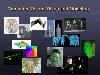
Vision • Vision is important for primates • ~50% of cortex devoted to visual perception • Stimulus in the visual system is light (electromagnetic energy) • We only see a small band of electromagnetic waves
The Eye • Retina • Photoreceptors • Rods – low levels of light • Cones – color • Bipolar cells • Ganglion cells
Form Eye to the CNS • Each eye is divided into two identical halves • Within each eye, one half received stimulation from the left visual field and the other from the right visual field • Optic nerve • Lateral or temporal branch stays on the same side • Medial or nasal branch crosses over • Optic chiasm
Form Eye to the CNS • Lateral geniculate nucleus (LGN) of the thalamus • Primary visual cortex (90%) • Geniculostriate pathway • Superior colliculus (10% - projects back to thalamus and then to cortex) – tectopulvinar pathway
Visual Cortex – Primary Visual Cortex • Different names for primary visual cortex: • Brodmann’s area 17 • V1 • primary visual cortex • striate cortex (“striped” cortex)
Retinal Topography and Cortical Blindness • Damage of the primary visual cortex causes blindness
Retinal Topography and Cortical Blindness • Hemianopia – loss of pattern vision in either the left or right visual field • Quadrantanopia –blindness in one quadrant of the visual field – damage to the optic tract, LGN or V1
Cortical Blindness and Consciousness • Case D.B. • Area around the right calcarine fissure was removed for treatment of angioma • Reported not seeing anything in the left visual field • Able of pointing out where the light was in the left visual field • Blindsight – residual visual abilities within a field defect in the absence of acknowledged awareness • Importance of subcortical visual pathways
~30 cortical visual areas with distinct functions Each visual area has a topographic representation of external space in the contralateral hemifield (however, these get ‘less’ topographic as we get further up in the system) Visual Areas of the Cortex Outside the Primary Visual Cortex
Two General Projections From the Primary Visual Cortex • Dorsal stream – Occipito-parietal stream spatial perception – action- “where” or “how to” • Ventral stream – Occipito-temporal stream object perception – identification – “what” stream
PET studies of “What” and “Where” pathways • Where task: did the objects remain in the same position? • What task: did objects change?
Area MT or V5 MOTION • Cells in area MT respond to movement but not color • For example, this particular neuron in this monkey’s V5 area responds best when stimulus moved down and to the left
Imaging Visual Areas in Humans • Semir Zeki – What part of the brain processes movement in the visual field in humans? • PET scans • Experimental condition: black-and-white collage set in motion • Control condition? • Motion – area V5 (MT)
Area V4COLOUR • Semir Zeki – What part of the brain processes colour in the visual field in humans? • PET scans • Experimental condition: multicolor rectangles • Control condition? • Colour – area V4
Deficits in Motion Perception: Akinetopsia • Case M.P. • Bilateral damage to teporolateral corticies (MT?). • “When I’m looking at the car first, it seems far away. But then when I want to cross the road, suddenly the car is very near.” • Color discrimination OK • Object recognition OK
Deficits in Color Perception - Achromatopsia • Congenital colorblindness (dichromats) vs. acquired colorblindness • Usually associated with damage to V4 • Colorblind painter – case J.I. • Object recognition OK • Improved acuity • People were “rat-colored” • Dreams?
“What” Pathway • Early in the stream, the cells are responsive to simple stimuli • Further up the stream, cells respond to more complex stimuli • Receptive field of a cell is the area of visual space to which the cell is sensitive • Cells in the primary visual cortex have small receptive fields • Cells further down the “What” stream have large receptive fields
Deficits Following Damage to the WHAT Pathway • Visual agnosia – partial or total inability to recognize visual stimuli, unexplainable by a defect in elementary sensation or reduced level of alertness or memory • NO GENERAL LOSS OF KNOWLEDGE • Different from other neurological conditions such as Alzheimer’s disease • Tactile agnosia (Asterognosis) • Auditory agnosia
Dissociating Deficits in WHAT and WHAT/HOW Pathways • Patient D.F. severe disorder of object recognition following carbon monoxide poisoning that produced damage in the lateral occipital cortex • Dissociating the “what” and “where” system • D.F. had no problem with the “where” pathway
Deficits Following Damage to the WHERE/HOW Pathway • Patient V.K. – bilateral hemorrhages in the occipitoparietal regions • Deficits with “how/where” stream but not “what” stream (can recognize objects • Optic ataxia – difficulty in using visual information to guide actions that cannot be ascribed to motor, somatosensory, or visual-field or –acuity deficits.
Subtypes of Visual Agnosia • Apperceptive agnosia • Associative agnosia
Apperceptive Agnosia • Apperceptive agnosia – is a fundamental difficulty in forming percept (a mental impression of something perceived by the senses) • cannot recognize, copy, or match objects, however elementary sensory functions appear relatively intact (i.e., patients are not blind). • usually bilateral damage to lateral portions of the occipital lobes (what stream – early deficits in visual perception) • Often associated with carbon monoxide poisoning
Associative Agnosia • Associative agnosia–basic visual information can be integrated to forma meaningful perceptual whole, yet that particular perceptual whole cannot be linked to stored knowledge • What is affected? “higher cognitive” level of processing that is associated with stored information about objects – that is with memory. • Patients have either lost access to memories of what things should look like or actually lost these memories • Damage to regions in ventral stream that are further up the processing hierarchy, such as the anterior temporal lobe
Associative Agnosia - Example • Case F.R.A. – infarct of the left posterior cerebral artery • Copying of objects OK • Can describe objects when they are named • Can segment a complex drawing into parts (apperceptive patients cannot do this) • He could not name these objects • How would test if this is a language problem (finding words for objects)?
Associative Agnosias Category Specificity • Patient J.B.R. • Herpes simplex encephalitis • Living objects (6% correct) • Inanimate objects (90% correct) • Other patients the other way around • Why is this the case? • Associative agnosia loss of semantic knowledge • Semantic knowledge has categories
Why Is It More Likely That Animals Won’t be Recognized? • Manufactured objects are manipulated • Associated with kinesthetic and motoric representations • Manufactured objects are easier to recognize because they activate additional forms of representations • Individuals can “where” or “how to” pathway to derive knowledge of an object might be
Patient C.K. • Using hand movements in order to identify an object
Special Category - Faces • Prosopagnosia is the inability to visually recognize familiar faces including their own • Can recognize people by their voice or birthmark or characteristic hairdo. • Prosopagnosia patients can be object-agnosia free • Are faces special? • Do the processes for faces and object involve physically distinct mechanism? • Are the systems (object and face recognition) functionally independent? • Double dissociation? • Do the two systems process information differently?
One Possibility- Hierarchical Model • If this model is correct, what would you expect regarding the dissociation between object and face recognition FACE PERCEPTION OBJECT RECOGNITION EARLY VISUAL PROCESSING
Dissociations of Face and Object Perception • Patient C.K. was presented with Giuseppe Arcimbaldo’s paintings • severe object agnosia but no prosopagnosia • Hierarchical model probably not correct • 2 parallel system (object recognition and face recognition) Perceives a face No perception
More Evidence that Faces are Processed Separately • Yin, 1970 • It is more difficult to process inverted faces • When inverted, faces are processed as objects • It is less difficult to recognize inverted objects
More Evidence that Faces are Processed Separately • Tanaka & Farah 1993 • Face perception not simply analysis of parts • For house perception it did not matter if the house was presented as a whole or in parts • Faces need to be perceived as whole
Are Faces Really Special or Are They Similar Examplars of the Same Category? • McNeil and Warrington, 1993 • Farmer with prosopagnosia • Tested on human faces – failed • Tested on sheep faces – OK • Is prosopagnosia due to the fact that faces are similar members of the same category?
Are Faces Really Special or Are They Similar Examplars of the Same Category? • Myles-Worsley, Johnson & Simons, 1988 • Experience influences the way items are processed
Are Faces Really Special or Are They Similar Examplars of the Same Category? • Bornstein, 1963 • Following brain damage, avid bird watcher could no longer distinguish between different bird species • Sergent & Signoret, 1992 • Toy car expert (5000 in his collection) • Following brain damage he became prosopagnosic • He was able to identify different cars
Neural Mechanisms for Face Perception • Martha Farah (1990) – looked at 71 prosopagnosic patients • Bilateral lesions – 65% • 29% right hemisphere lesions • 6% left hemisphere lesions lesions • Conclusions?
Neural Mechanisms for Face Perception • Single-cell recordings in monkeys • Various stimuli presented including monkey and human faces • Activation in the inferior temporal cortex
Neural Mechanisms for Face Perception in Humans • Human fMRI studies • Face stimuli associated with activation of the right fusiform gyrus • Fusiform face area – FFA • FFA is also activated by other stimuli • Car and bird experts
Activation of the FFA by Non-Facial Stimuli • Individuals were trained to be experts at recognizing “greebles” • In “greebles” experts, FFA is activated during identification