Download
1 / 28
320 likes | 981 Views
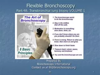
Prepared By Bronchoscopy International Contact us at BI@bronchoscopy.org. Flexible Bronchoscopy Part 4B : Transbronchial Lung Biopsy VOLUME 2. Response to procedure-related complications and adverse events. Transbronchial lung biopsy (TBLB) Volume 2. AIRWAY BLEEDING And PNEUMOTHORAX.

E N D
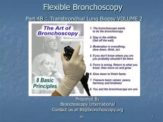
Prepared By Bronchoscopy International Contact us at BI@bronchoscopy.org Flexible BronchoscopyPart 4B :Transbronchial Lung Biopsy VOLUME 2 BI
Response to procedure-related complications and adverse events Transbronchial lung biopsy (TBLB) Volume 2 AIRWAY BLEEDING And PNEUMOTHORAX Bronchoscopy International
Generally reported frequency of complications after Transbronchial lung biopsy • Bleeding > 50 ml 1-2 % • Pneumothorax 1-4 % • Death 0.04 - 0.12 % BI
Bleeding after biopsy • Increased risk in case of • Coagulopathy • Platelet dysfunction • Platelets < 50,000 • Uremia • Immunocompromised host • Anticoagulation medication including certain antiplatelet medications such as Plavix • Increased risk suspected but not documented in • Congestive heart failure • Pulmonary hypertension BI
Prevention • Screening before airway procedures • History, examination, laboratory tests, explanation of risks to patient and or family members • Careful procedure technique • Recognize hypervascularization, aberrant vessels, and submucosal arterioles • Procedural planning • Supplemental oxygen, cardiac monitoring • Be sure sufficient space in procedure room to move around. • Availability of medication and hemodynamic resuscitation, including crash cart. • Airway resuscitation including endotracheal tubes, large bore suction catheter/Yankauer, oral airway and bite block. BI
Accepted precautions to prevent bleeding • Platelet counts > 50,000/mm3 • Avoid uremia (serum creatinine < 2, BUN < 25 mg/dl) • Avoid liver failure (alk phos < 110, SGOT < 25, Bilirubin < 1.5 ml/dl • Avoid anticoagulated patients • Check PT, aPTT in patients with history of bleeding or coagulopathy. • Stop antiplatelet agents such as Plavix BI
Morbidity related to • Physiologic consequences of airway bleeding • Blood filling of dead-space • Airway obstruction and clot formation • Subsequent tachypnea and hypoxemia • Tachycardia, bradycardia, hypotension • Respiratory failure • Arrhythmia and cardiac arrest • Underlying disease state • History of pneumonectomy • Critically illness • Significant comorbidities BI
Bronchial arterial anatomy • Bronchial arterial blood (systemic arterial pressures) • Comes from the aorta (T 3-T 8) • Feeds the trachea and main bronchi • Drains into the bronchial veins and right heart • Feeds intrapulmonary tissues and airways • Drains through bronchopulmonary anastomoses into pulmonary veins and left heart Collateral circulation and increased bronchial and pulmonary anastomoses are found in inflammatory diseases, cystic fibrosis, bronchiectasis, and TB. BI
Left upper lobe pulmonary veins Left upper lobe pulmonary artery Vascular and airway anatomy Carina Left Pulmonary artery Main pulmonary artery BI
Ventilatory dead space A patient’s left main bronchus, right main bronchus, and trachea can completely fill with only 150 ml of blood or saline, causing hypoxemia, and respiratory arrest. BI
Treating the bleeding airway • Establish and maintain an open airway • Stop the bleeding • Prevent or treat respiratory, cardiac, and hemodynamic complications BI
(1) Maintaining an open airway • Bronchoscopic suction and large bore suction of the oral pharynx • Lateral safety position • Tilt the patient or the table 45 degrees towards the bleeding side • Note the bleeding site and remember how to get back to it! • Tamponade the bleeding bronchus using continuous bronchoscopic suction • Unilateral intubation BI
The safety position (lateral decubitus) • Bleeding side down • Allows face to face contact with patient if operator working from the front or side of the patient • Allows blood and secretions to flow from the larynx and out of the corner of the mouth • Avoids collapse of the larynx and laryngeal obstruction by tongue or edematous upper airway. • Oral pharynx easily suctioned BI
Safety position Turning the patient onto the “safety position” (bleeding side down) also protects the contra lateral airway BI
(2) Stop the bleeding • Tamponade using • Bronchoscopic suction, Balloons, the rigid bronchoscope, cotton pledgets, tampons. • Vasoconstriction using • Epinephrine, cold saline washes • Intravenous vasopressin (0.2 - 0.4 units / min) causes bronchial arterial vasoconstriction: danger if patient has coronary artery disease and hypertension. • Enhance clot formation • Allow clot to form in the bleeding area • Lateral decubitus position BI
Tamponade balloons If a tamponade balloon or Fogarty catheter is inserted into a bleeding segmental bronchus, its position should be verified by flexible bronchoscopy and chest radiograph. The balloon can remain in place for several days if necessary. BI
Dilating balloons Tamponade balloons or, if necessary, dilating balloons are usually large enough to tamponade a bleeding segmental and subsegmental airway BI
Fogarty catheters A Fogarty balloon catheter can be used but operators and their assistants should first verify that balloon diameter is sufficient to fill segmental bronchial airway AND that balloon catheter fits through working channel of the bronchoscope. BI
The Cook (Arndt) bronchial blocker, if necessary, should be inserted through a large endotracheal tube BI
Saline lavage Immediate administration of large aliquots of iced saline using a wedged or partially wedged bronchoscope and continuous or intermittent suction and gravity dependent clot formation stops most bleeding. BI
Do not remove freshly formed clot Once a clot forms, it is important to NOT remove it once bleeding has stopped. Inspection bronchoscopy (with or without clot removal can be performed the following day Large blood clot causing a cast of the distal airway BI
Avoid adverse effects on respiration , cardiac, and hemodynamic status: Beware anxiolytics and narcotics on respiration In case of bleeding, additional intravenous sedation can result in adverse events: These include respiratory failure, hypoxemia, and hypercapnia, hypotension and aspiration pneumonia. Reversing agents should be available. Additional sedation or anxiolysis might warrant intubation even after bleeding is controlled. BI
Avoid adverse effects on respiration , cardiac, and hemodynamic status: Consider intubation with a large endotracheal tube If intubation is desired or warranted, a large single lumen endotracheal tube can usually be inserted over the bronchoscope. Selective unilateral bronchial intubation is only possible if the oral route is used. ALWAYS insert a bite block to prevent patients from biting down on the bronchoscope (regardless of level of sedation). BI
Pneumothorax after biopsy • May be immediate • Detected by symptoms such as dyspnea, pleuritic chest pain, hemoptysis, tachycardia, tachypnea, or hypotension. • Detected on fluoroscopy • May also be delayed • Justifies prolonged observation post-procedure • May be detected by symptoms, or chest radiograph (during exhalation) • May often be small and asymptomatic BI
Treatment alternatives • Observation and repeat chest radiograph if small and asymptomatic. • Observation and hospital admission. • Small bore chest tube insertion and discharge. • Small bore chest tube insertion and hospital admission. • Large bore chest tube insertion and hospital admission. BI
Examples of chest tubes A B A Pigtail B. Cook catheter C. Tru-Close D. One-way valve C D BI
This presentation is part of a comprehensive curriculum for Flexible Bronchoscopy. Our goals are to help health care workers become better at what they do, and to decrease the burden of procedure-related training on patients. BI
Bronchoscopy International: Art of Bronchoscopy, an Electronic On-Line Multimedia Slide Presentation. http://www.Bronchoscopy.org/Art of Bronchoscopy/htm. Published 2007 (Please add “Date Accessed”). All efforts are made by Bronchoscopy International to maintain currency of online information. All published multimedia slide shows, streaming videos, and essays can be cited for reference as: Thank you BI