Download
1 / 14
140 likes | 602 Views
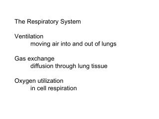
Air Into the Lungs. Lungs and Respiratory Tree Lungs and Pleural Cavity Diaphragm Costal muscles Lung Volume and Respiratory Control. Alveoli reminder—this is where the action is!. Key to lung function Where O2 enters blood, CO2 leaves blood Every alveoli is capillary covered sac

E N D
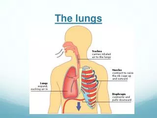
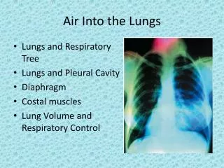
Air Into the Lungs • Lungs and Respiratory Tree • Lungs and Pleural Cavity • Diaphragm • Costal muscles • Lung Volume and Respiratory Control
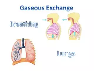
Alveoli reminder—this is where the action is! • Key to lung function • Where O2 enters blood, CO2 leaves blood • Every alveoli is capillary covered sac • Lining of sac is squamous epithelium • Also cuboidal epithelial cells that secret surfactant (keeps surfaces from sticking) and cilia (to move mucous and particles up respiratory tree • Unfold alveolar membranes in human lungs—tennis court! But how does air get to alveoli?
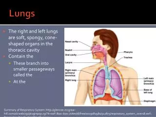
Lungs and Respiratory Tree • Air enters through nasal passage to pharynx to glottis (A and P I review) • Trachea to primary bronchii (left, right) to secondary bronchii to tertiary bronchii to bronchioles to clusters of alveolii in pulmonary lobules • Cartilage rings hold trachea and bronchii open • NAV (Nerve, Artery, Vein) bundle follow branching bronchii to capillary beds that surround alveoli
Respiratory epithelium • Bronchii lined by ciliated, pseudo-stratified epithelium • Goblet cells and glands secrete mucous • Cilia move in waves and mucous, along with particles are swept towards pharynx—”mucous escalator”
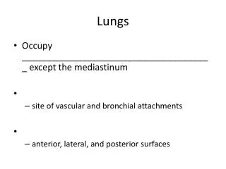
Lungs and Pleural cavity—visceral and parietal pleura • Serous linings of body cavity (A and P I review)—lubricated to prevent sticking • Parietal pleura on internal surface or ribs/costal muscles • Visceral pleura adhered to external surface of lungs • Pleural cavity is between visceral and parietal pleura which seal the cavity
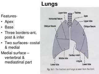
Lobes of lungs • Right lung • Upper lobe • Middle lobe • Lower lobe • Left Lung • Upper lobe • Lower lobe • Cardiac notch
Action of the Diaphragm • Primary muscle of respiration (involuntary) • Contraction during inspiration • Increases volume of thoracic cavity • Decreases pressure of thoracic cavity • Air moves into lungs (highlow pressure) • Forced contraction (voluntary) • Used for defecation, urination, labor • Increases pressure in abdominal cavity • Pushes on abdominal organs to move contents out Diaphragm can only actively contract inferiorly to expand thoracic cavity. Exhalation of air from lungs is due to elastic tissue in lungs and compression of abdomen by abdominal muscles
Liver as Piston During running, in humans and quadrupedal mammals, the large, heavy liver, moves cranially and caudally, due to rhythmic momentum changes as the feet hit the ground. This in turn pushes and pulls on the diaphragm, thereby expanding and contracting the thoracic cavity with little muscular action on the part of the diaphragm itself.
Other respiratory muscles • External intercostals work with diaphragm to help expand rib cage during inhalation • Internal intercostal muscles with abdominals can force contraction of rib cage during exhalation.
Rate of air exchange • At rest: 500 ml/breath (tidal volume) x 12 breaths/min (resting respiratory rate) = 6 liters/min. • Peak exercise: 4800 ml/breath (expanded tidal volume using most of reserves) x 50 breaths/min. (increased respiratory rate) = 200 liters/min. Lung Volume and Respiratory Rate (review) Lung Capacity • Total lung capacity: 4200 ml (f); 6000 ml (m) • Resting tidal volume: 500 ml (f) • Expiratory reserve (amount you can blow out after exhaling normally): 700 ml (f) • Residual volume (amount you can never blow out): 1100 ml (f) • Inspiratory reserve (amount you can breathe in after normal inhalation): 1900 ml (f)
Respiratory control (review) • Great review of nervous system • Brainstem • Cranial nerves • Sensory and motor • Visceral versus somatic • CO2 levels have more input during normal respiration • Possible to blow off enough CO2 to pass out since low O2 doesn’t kick in on time—careful divers and swimmers!