The Integumentary System
310 likes | 342 Views
Explore the organs and layers of the integumentary system, including the epidermis, dermis, and subcutaneous layers. Learn about the functions of the skin, such as protection, temperature regulation, and sensory reception. Discover common skin conditions and their treatments, as well as the role of the integumentary system in synthesizing vitamin D and the prevention of skin cancer.

The Integumentary System
E N D
Presentation Transcript
Organs Two or more types of tissues grouped together, perform specialized functions Largest organ?
Functions Protective covering Regulates body temp Prevents water loss Sensory receptors Makes biomolecules Excretes waste Makes vitamin D
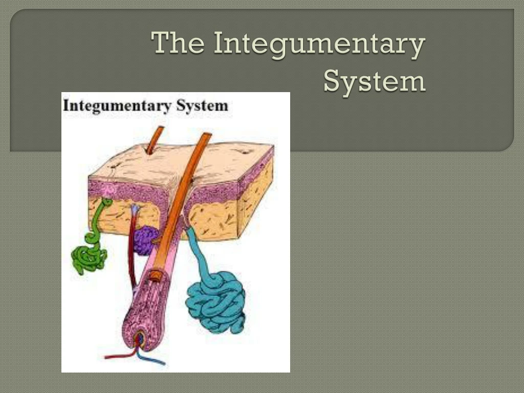
Skin Layers • Epidermis, dermis, subcutaneous (hypodermis)
Epidermis • Outer layer • Thin • Made of stratified squamous epithelial cells (no blood vessels) • Constant cell division pushes older cells upwards. Cells die off in 2-4 weeks • Keratin (protein) enters and hardens • Palms, soles of feet have lots!!
Skin Colors • Skin color variations result from proteins in the epidermis • Melanin • Carotene • Hemoglobin • Bilirubin
Skin Colors • Melanin: produced by melanocytes • Melanocytes about equal in all people • Greatermelanin results in darker skin color • Absence of melanin referred to as albinism
Skin Color • Carotene • from beta-carotene • Gives skin an orange-yellowish color • Color lightens as carotene breaks down
Skin Color • Hemoglobin • Protein in blood • Capillaries in skin dilate allowing more blood to flow to surface • Gives skin a pinkish/reddish appearance • Goes away when capillaries contract
Skin Colors Bilirubin Builds up during jaundice, turns skin yellow
Dermis • Below epidermis • Thicker • Dense connective tissue • Has projections into epidermis to anchor it – causes spiraling patterns • fingerprints
Dermis The dermis contains: • nerve fibers • sensory fibers • hair follicles • sebaceous glands • sweat glands • blood vessels
Subcutaneous (hypodermis) • Belowdermis • Binds skin to underlying organs • Mostly adipose tissue • Provides protection from shock • Insulation • Blood Vessels *Not a true skin layer
Bedsores/Pressure Ulcers -Interference with blood flow to the dermis can killepidermal cells -Lying in one position too long causes weight of body to block skin’s blood supply -Treatment: shift patient, wound cleaning, massage
Accessory Organs • Hair • Grows from hair follicle • Arrectorpilimuscle makes hair “stand up” when cold • Dead, keratinized, epidermal cells
Accessory Organs • Nails stratified squamous, keratinized, epithelial cells overlying the nail bed the lunulais the most actively growing region of the nail root – (white section at proximal end of nail)
Accessory Organs Sebaceous Glands • Secretesebum (oil) into hair follicle • Lubricates and waterproofs hair and skin • Bacteria in follicle produces red pimple
Accessory Organs • Sweat Glands • Two types -Eccrine: excrete sweat onto skin; respond primarily to body temp -Apocrine: excrete sweat into hair; respond primarily to stress, also body temp and sexual arousal (become active during puberty)
Hyperhidrosis • Overactive sweat glands -Hands -Feet -Armpits • Caused by overactive nervous system • Treatment: Antiperspirant Iontophoresis Botox injections
Skin Functions • Protection • Sensory reception- specialized cells embedded in skin detect hot, cold, pain, touch • Body temperature regulation –changes in blood vessel diameter and sweat gland production for hot/cold
Skin Functions Synthesis of Vitamin D • Molecules (dehydrocholesterol) exposed to UV rays turn into vitamin D • Vitamin D converts into a hormone called calcitronin in kidneys • Regulates calcium and phosphate levels • Prevents rickets • Promotes bone health
Skin Cancer-Carcinomas • Non-pigmented epithelial cells • More common, slow growing • Light skinned people, over 40 years • Hard, dry, scaly growths • Usually surgicallytreated or with radiation
Skin Cancer- Melanoma • Malignant Melanoma • Serious- can lead to death • Resembles a mole – dark spot • Can be caused, by short, intermittent exposure to high intensity sunlight • Any age • First grows horizontal (surgically removed), then downward and can spread into deeper tissues
Skin Burn Severity • 1st degree: only epidermis is affected • 2nd degree: all of epidermis and part of dermis affected • 3rd degree: all of the epidermis and dermis are destroyed • 4th degree: reaches muscle or bone • Can go up to 5th degree
Rule of the Nines • Body is divided into 11 sections • Each section takes about 9% of body’s skin to cover it • Add up all areas of body that are burned badly enough to blister • Used in the field to determine where to take patient for treatment
Types of Injections • Subcutaneous: into subcutaneous layer; • vaccines/flu shot • insulin, • morphine, • penicillin • Intradermal: into the dermis • Local/regional anesthetics, • allergy tests • TB test
Injections • Intramuscular: into the muscle • Quick absorbtion; • antibiotics, • hormones, • codeine, • epinephrine, • Botox • Intravenous: into a vein • Fluids, • blood transfusions, • lethal injections


