Download
1 / 21
250 likes | 590 Views
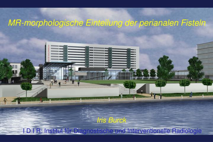
MR-morphologische Einteilung der perianalen Fisteln. Iris Burck I D I R: Institut für Diagnostische und Interventionelle Radiologie. Perianale Fisteln. Ätiologie: Abflussstörung und Infekt der Proktodealdrüsen (Abszess- oder Fistelung)

E N D
MR-morphologische Einteilung der perianalen Fisteln Iris Burck I D I R: Institut für Diagnostische und Interventionelle Radiologie
Perianale Fisteln Ätiologie: Abflussstörung und Infekt der Proktodealdrüsen (Abszess- oder Fistelung) Entzündliche Fortleitung bei Appendizitis, Divertikultis Morbus Crohn, Colitis ulcerosa (Ulzeration der Rektummukosa) Geburtstrauma (anorektale-vaginale Fisteln) Strahlentherapie Epidemiologie: M >w , Altersgipfel 30.-40. LJ, 2% Life- time-risk: intersphinktere oder transphinktere Fisteln ca. 95% der Fälle Hippokrates von Kos Institut für Diagnostische und Interventionelle Radiologie, J.W. Goethe-Universität, Frankfurt
IRF = ischorektale Fossa IAF = ischioanale Fossa Institut für Diagnostische und Interventionelle Radiologie, J.W. Goethe-Universität, Frankfurt
„Anal clock“ SSL = Stein-Schnitt-Lage ….surgeons and radiologists with the same point of view! Institut für Diagnostische und Interventionelle Radiologie, J.W. Goethe-Universität, Frankfurt
Goodsall-Regel: Fistelöffnungen cranial des Analhorizontes verlaufen gradlinig, Fistelöffnungen caudal verlaufen bogenförmig und münden meist bei 6Uhr SSL im Analkanal Proktodealdrüsen: rudimentäre „Duftdrüsen“, intersphinkter gelegen, meist an der hinteren Kommissur (=dorsal), meist 6 Uhr SSL in ca.80% Institut für Diagnostische und Interventionelle Radiologie, J.W. Goethe-Universität, Frankfurt
Normale Anatomie Institut für Diagnostische und Interventionelle Radiologie, J.W. Goethe-Universität, Frankfurt
Entnommen aus: Morris J et al. (2000) MR imaging classification of perianal fistulas and its implications for patient management. RadioGraphics 20:623-635 Institut für Diagnostische und Interventionelle Radiologie, J.W. Goethe-Universität, Frankfurt
Grad 1 - intersphinkter Institut für Diagnostische und Interventionelle Radiologie, J.W. Goethe-Universität, Frankfurt
Grad 2 – interspinkter mit Abszess Institut für Diagnostische und Interventionelle Radiologie, J.W. Goethe-Universität, Frankfurt
Grad 2 - Hufenheisen-Typ - intersphinkter Institut für Diagnostische und Interventionelle Radiologie, J.W. Goethe-Universität, Frankfurt
Grad 3 - transsphinkter Institut für Diagnostische und Interventionelle Radiologie, J.W. Goethe-Universität, Frankfurt
Grad 4 – transpinkter mit Abszess Institut für Diagnostische und Interventionelle Radiologie, J.W. Goethe-Universität, Frankfurt
Grad 4 – transsphinkter mit Abszess Institut für Diagnostische und Interventionelle Radiologie, J.W. Goethe-Universität, Frankfurt
Grad 5 – supralevatorisch / translevatorisch Institut für Diagnostische und Interventionelle Radiologie, J.W. Goethe-Universität, Frankfurt
Überblick Grad 3 Grad 1 Grad 2 Grad 5 Grad 4 Institut für Diagnostische und Interventionelle Radiologie, J.W. Goethe-Universität, Frankfurt
Alternative Bildgebung • Fisteldarstellung unter Durchleuchtung • Probleme: Sphinkterkomplex-Darstellung und genaue topographische Lage der Fistel • Endosonographie • Probleme: Externer Sphinkter schwer darstellbar, DD Fibrose versus Infektion, ungenügende Eindringtiefe (Begleitprozesse) • CT mit i.v. und rektaler KM-Gabe • Probleme: anatomische Differenzierung (Sphinkterkomplex, Levator ani), DD Fibrose versus Infektion, Artefakte durch KM-Leckage oder Gaseinschlüsse Institut für Diagnostische und Interventionelle Radiologie, J.W. Goethe-Universität, Frankfurt
CASE: 46jähriger Patient mit V. a. glutealen Abszess. CRP 27 mg/dl, Leukozyten 14.000/nl. Grad 5 mit transphinkterer Fistelung Institut für Diagnostische und Interventionelle Radiologie, J.W. Goethe-Universität, Frankfurt
Kontinenzorgan • M. sphinkter ani internus: 85% des Ruhetonus, kann durchtrennt werden meist ohne Kontinenzverlust • M. puborectalis • M. levator ani • M. sphinkter ani externus: 15% des Ruhetonus, sehr vulnerabel • „if this ring be cut, loss of control surely follows…“ Institut für Diagnostische und Interventionelle Radiologie, J.W. Goethe-Universität, Frankfurt
Quellenangaben: • Atlas der Anatomie Frank H. Netter Urban&Fischer 4.Auflage, 2008 • Morris J et al. (2000) MR imaging classification of perianal fistulas and its implications for patient management. RadioGraphics 20:623-635 Institut für Diagnostische und Interventionelle Radiologie, J.W. Goethe-Universität, Frankfurt
Institut für Diagnostische und Interventionelle Radiologie, J.W. Goethe-Universität, Frankfurt
FISI 2008 9. Frankfurter Interdisziplinäres Symposium für Innovative Diagnostik und Therapie MRT-Diagnostik Bewährtes und neue Entwicklungen Frankfurt/Main, 26. - 27. September 2008 www.fisi-frankfurt.de