IMPROVED MRI TEMPERATURE IMAGING USING A SUBJECT-SPECIFIC BIOPHYSICAL MODEL
350 likes | 675 Views
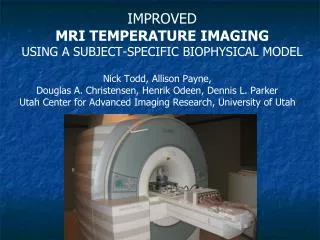
IMPROVED MRI TEMPERATURE IMAGING USING A SUBJECT-SPECIFIC BIOPHYSICAL MODEL . Nick Todd, Allison Payne, Douglas A. Christensen, Henrik Odeen, Dennis L. Parker Utah Center for Advanced Imaging Research, University of Utah . Background. Utah Projects in MRI guided HIFU

IMPROVED MRI TEMPERATURE IMAGING USING A SUBJECT-SPECIFIC BIOPHYSICAL MODEL
E N D
Presentation Transcript
IMPROVEDMRI TEMPERATURE IMAGING USING A SUBJECT-SPECIFIC BIOPHYSICAL MODEL Nick Todd, Allison Payne, Douglas A. Christensen, Henrik Odeen, Dennis L. Parker Utah Center for Advanced Imaging Research, University of Utah
Background • Utah Projects in MRI guided HIFU • Large animal MRgHIFU system (Siemens/IGT) • Small animal MRgHIFU system (IGT) • Breast MRgHIFU system • (UofU/IGT/Siemens) • See poster 4.8 by Allison Payne
Background: Utah Projects MRI scanner Tumor • MR guided HIFU • Breast: • Develop the Utah Breast MRgHIFU system • Brain • Develop 3D MRI Temperature measurements for MRI guided Brain HIFU • Temperature measurement requirements • Breast: • glandular tissues AND fat • Near-field protection • Brain: • cover entire skull volume • high temporal and spatial resolution
MR Temperature Basics Proton Resonance Frequency Shift (PRF). MR signal frequency depends on local chemical environment of water Hydrogen. Temperature changes affect this environment. Current Time Frame Reference Difference Temperature Map Frequency changes measured as image phase changes. - =
Breast: Temperature Measurements 3-point Dixon Images • Requirements: • Control treatment in glandular tissue • Avoid fat necrosis • Coverage, speed, and resolution • Temperature in water and fat? • Hybrid PRF/T1 method • 2D GRE • 2D/3D Segmented EPI Fat Water fat agar PRF temperature map T1 signal change map
MRI Thermometry - Breast Hybrid PRF/T1 Signal from Spoiled GRE sequence: Image sequence: 2 alternating flip angles PRF from phase of each image T1 from two images Deoni, Rutt, Peters. Magn Reson Med 2003 49:515-526.
Breast: Temperature Measurements A) 3-Pt Dixon Water Image B) 3-Pt Dixon Fat Image C) PRF/T1 Magnitude Image D) PRF Temperature Map Pork Muscle Breast Fat Targeted Area Transducer
Breast: Temperature Measurements T1 Percent Change in Breast Fat PRF Temperatures in Pork B) C) PRF/T1 Magnitude Image D) PRF Temperature Map Pork Muscle Breast Fat Targeted Area Transducer
Transcranial MRI guided HIFU Funding: Focused Ultrasound Surgery Foundation NIH R01 EB013433
Transcranial MRI guided HIFU • Cover all heated regions: Skull + within • Resolution Speed Coverage (FOV) • 1mm isotropic 1s Full head/breast • 205x160x100 TR=35, ETL=7: 80s • 205x160x33 (1x1x3mm), TR=35, ETL=7: 27s Image Volume
Required Values 1 x 1 x 3 mm Spatial Resolution: Temporal Resolution: Volume Coverage: Signal - to - Noise: 2 seconds per image 256 x 162 x 72 mm Brain Image Volume: 256 x 162 x 72 mm Image Volume
Transcranial MRI guided HIFU • How to go faster: • 2D Spatially selective RF excitation • Prefer full FOV • Parallel imaging + UNFOLD1 • Temporally Constrained Reconstruction (TCR)2 • Model Predictive Filtering (MPF)3 • 1: Chang-Sheng Mei, et al. Magnetic Resonance in Medicine 66:112–122 (2011) • 2: N. Todd et al. Magn Reson Med 62(2):406-419 (2009). • 3: N. Todd, A. Payne, D. L. Parker, Magn Reson Med 63:1269–1279 (2010)
Data Acquisition & Reconstruction F = Fourier Transform m = Image Estimate d’= Undersampled Data ~ Data Space (k-space) Image Space Inverse Fourier Transform 256 x 162 x 24 pixels 256 x 162 x 24 pixels
Constrained Reconstruction F = Fourier Transform m = Image Estimate d’= Undersampled Data = Gradient in time ~ is iteratively updated subject to constraints: Image must match acquired data Image must change smoothly in time iteration = 5 iteration = 25 iteration = 50 iteration = 100
TCR: Constrained Reconstruction • Sequence Parameters • 1.5 x 2 x 3 mm • 288 x 216 x 108 mm • 192 x 108 x 36 matrix • EPI Factor: 7 lines per excitation • TR/TE = 35 / 9 ms Data Undersampling ky kz Constrained Reconstruction Scan Time: 1.8 s / time frame 25 s / full data set Not real time
Constrained Reconstruction Results Validation Tests: “Truth”: Full Data used 1.5 x 1.5 x 3.0 mm 2.8 seconds per image 288 x 162 x 24 mm Test Cases: 288 x 162 x 48 mm 288 x 162 x 90 mm 288 x 162 x 144 mm 2.8 s Full Data “Truth” 5.4 s 10.1 s 16.2 s Constrained Reconstruction 6X data reduction 2.8 s “Truth” 0.9 s 1.7 s 2.7 s
Model Predictive Filtering Thermal Model Artifact-free Temperature maps Undersampled k-space Goal: real time N. Todd, A. Payne, D. L. Parker, MRM 63:1269–1279 (2010)
Model-Predictive Filtering • Segment tissues • Determine tissue-specific thermal and acoustic properties • TCR + Modeling • Use tissue-specific properties in dynamic MPF temperature measurements • Realtime, 3D, large FOV • From highly undersampled 3D segmented EPI PRF
Tissue Segmentation • Breast tissue segmentation • Hierarchal Support Vector Machine algorithm 3pt Dixon H2O only FS PD-w 3pt Dixon Fat only Non-FS T1 FS T2-w h-SVM w/ Zero-Filled-Interpolation
Tissue property estimation:Acoustic parameters Segment treatment volume into a small number of tissue types 4-8 low power pulses cover targeted volume TCR – reconstruct temperature images In-vivo estimates of the change in the attenuation coefficient with log10 of thermal dose using the iterative parameter estimation technique . Urvi Vyas et al. ISTU 2011 MR temps to get SAR patterns Use ultrasound model (HAS) to determine absorption and speed of sound to match measured pattern Tissue acoustic values for Model Predictive Filtering.
Tissue property estimation:Thermal parameters Segment treatment volume into a small number of tissue types 4-8 low power pulses cover targeted volume TCR – reconstruct temperature images MRI temps during cooling Determine thermal diffusivity using cooling temperature curves Cheng et al., JMRI 16(5), 2002
HAS: Head Model Courtesy: Guido Gerig, University of Utah
Model Predictive Filtering Multi-step, recursive algorithm 3 2 Phase (n+1) 1 3 Temp (n+1, model) Temp (n) K-space (n+1) 5 Magnitude (n) Step 1: Use model to predict temperature at time n+1. Step 2: Convert temperature map to phase map for time n+1. Step 3: Use this phase and the magnitude from time n to create k-space for time n+1. Step 4: Insert any actually acquired k-space lines. Step 5: Recalculate the temperature for time n+1 using the data updated k-space. Temp (n+1, model and data)
Model Predictive Filtering Use the Pennes Bioheat Equation, tissue properties, and a pre-treatment heating to determine the thermal model. Full Data Model Only T = temperature r = density C = tissue and blood heat capacity k = thermal conductivity Wb = blood perfusion Q = heat applied
2-D MPF Results Fully sampled k-space data sets: 288x288x20mm FOV, 2.3x2.3x4mm res, 8.3 sec/scan. 25% of k-space used in reconstruction. Power = 36W (Model Id data set) Mean and STD of error over an ROI MPF Power = 42W Mean and STD of error over an ROI MPF Power = 48W Mean and STD of error over an ROI MPF
3D (R=12) vs 2D (R=1) MPF Temperatures Common: Ultrasound pulse = 36 W/58.1 sec 3-D GRE: FOV = 256x256x32 mm3, Matrix = 128x128x16 Resolution = 2.0x2.0x2.0 mm3 TR/TE = 25/8 ms Tacq = 76.8 s/image volume (R=1) = 6.4 s/image volume (R=12.1) 2D GRE: FOV = 256x256x20 mm (sl = 3mm) Matrix = 128x128 Resolution = 2.0x2.0x3.0 mm3 TR/TE = 65/8 ms; 8.3 sec per scan (R=1) Scans repeated 8x for variability N. Todd, A. Payne, D. L. Parker, MRM 63:1269–1279 (2010)
Model Predictive Filtering Results Phantom Heating 2.0 x 2.0 x 2.0 mm 0.5 seconds per image 256 x 162 x 48 mm σT< 1°C Transverse: Sagital: Coronal:
Summary: Work in Progress • Brain requires: • Large FOV: Cover insonified volume • High speed: 1s/volume • High resolution: < 1 x 1 x 3 mm3 • Our solutions: • PRF: Highly undersampled (>8) 3D segmented EPI • TCR: • Does not require tissue thermal and acoustic properties • Achieves high spatial and temporal resolution, large FOV, LOW NOISE! • Cannot (yet) be performed in real time • Model-predictive Filtering (MPF) • Requires • tissue segmentation • estimate of tissue acoustic and thermal properties • Property estimates: • SAR: Hybrid Angular Spectrum (HAS) • Diffusivity/Perfusion: MRI during cooling • Also achieves high spatial and temporal resolution, large FOV, LOW NOISE! • Potential real time application • Parallel imaging • Can be used to supplement TCR or MPF • Difficult with currently used HIFU coils
Acknowledgments Thank You People: Dennis Parker Bob Roemer Doug Christensen Leigh Neumayer Allison Payne Nick Todd Rock Hadley Nelly Volland MahamadouDiakite Yi Wang Urvi Vyas Emilee Minalga Joshua de Bever Chris Dillon Joshua Coon Justin Tidwell LexiFarrer Robb Merrill Henrik Odeen Funding: Focused Ultrasound Surgery Foundation Siemens Medical Solutions NIH grants F31 EB007892-01A1, R01 EB013433, and R01 CA134599.