Describing the Abdomen
480 likes | 1.02k Views
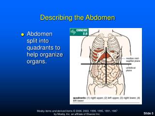
Describing the Abdomen. Abdomen split into quadrants to help organize organs. Describing the Abdomen. The abdomen can also be split into 9 sections as well. Right Upper Quadrant (RUQ). Liver & Gallbladder Head of Pancreas Duodenum Right adrenal gland Hepatic flexure of colon

Describing the Abdomen
E N D
Presentation Transcript
Describing the Abdomen • Abdomen split into quadrants to help organize organs.
Describing the Abdomen • The abdomen can also be split into 9 sections as well.
Right Upper Quadrant (RUQ) • Liver & Gallbladder • Head of Pancreas • Duodenum • Right adrenal gland • Hepatic flexure of colon • Portion of right kidney
Right Lower Quadrant (RLQ) • Appendix • Ascending colon • Ovary • Right ureter • Bladder • Uterus
Left Upper Quadrant (LUQ) • Spleen • Stomach • Pancreas • Left lobe of liver • Splenic flexure of colon • Portions of transverse and descending colon
Left Lower Quadrant (LLQ) • Lower left kidney • Sigmoid colon • Ovary • Left ureter • Descending colon • Bladder • Uterus
Assessing the Abdomen • Chief complaint • History of present illness • Past medical history • Family history • Personal/Social history • Physical Exam • Differential • Labs
Importance of History Present Illness • GI versus cardiac causes • Clues to Differential • Abdominal “Review of Systems” • Seven attributes of a symptom
Review of Systems for the Abdomen • Constipation • Jaundice • Dysuria • Urinary frequency • Hematuria • Abdominal pain • Indigestion • Nausea • Vomiting • Diarrhea
• Review of Related History Past Medical History • Gastrointestinal disorders • Urinary Tract Infections • Surgeries • Medications/Immunizations • Trauma/Injury • Blood transfusions • Hepatitis • Cancer
• Review of Related History Family History • Familial Mediterranean Fever • Inflammatory bowel disease • Familial cancer syndromes • Congenital malformations • Cystic fibrosis, Celiac’s • Kidney disease • Gallbladder disease
• Review of Related History Personal and Social History • Nutrition • LMP (first day of last menstrual period) • Stressful life events • Travel /Exposure to infectious diseases • Sexual history • Use of alcohol and/or illicit drugs • Tobacco use
• Review of Related History Pregnant Women • Urinary symptoms • Frequency, urgency, burning, suprapubic pain • Back pain • Abdominal pain • Contractions • Onset, frequency, duration, intensity • Leakage of fluid, blood
• Review of Related History Older Adults • Urinary Tract Infections • Constipation • Indigestion • Dietary habits
Abdominal Exam • Preparation • Inspection • Auscultation • Percussion • Palpation
Preparation for the Abdominal Exam • Empty bladder • Positioning • Abdomen exposure • Visualize anatomy • Warm hands & stethoscope • Approach from the right • Remember chief complaint • Watch face
Inspection (1) • Skin characteristics: color, striae, rashes, lesions, scars, dilated veins • Umbilicus: location, displacement, inflammation • Contour: rounded, flat, scaphoid • Symmetry: symmetric, bulges, distension • Surface Motion: pulsations, peristalsis
Auscultation • Bowel sounds: frequency, character • Vascular sounds: bruits, venous hums
Percussion (1) • Assess size & density of organs • Detect presence of fluid, air, masses, tenderness • Can be done independently or concurrently with palpation
Percussion (2) • All quadrants: tympany vs dullness • Liver span: at right MCL and at the MSL if enlarged • Spleen percussion sign: lowest left ICS before & after deep breath • Gastric bubble: left lower anterior rib cage, left epigastric area • Kidneys: costovertebral angle
Percussion (3) • If suspect ascites as result of percussion of abdominal wall may perform two additional tests: Shifting Dullness Test and the Fluid Wave Test
Palpation (1) • Evaluation of organs for: size, shape, consistency, tenderness • Evaluate umbilical ring • Palpate for pulsation of abdominal aorta • Detection of masses
Palpation (2) • Light Palpation: all 4 quadrants; identify muscular resistance, tenderness, masses • Deep palpation: all 4 quadrants; delineate organs and detect deeper masses • Mass: location, size, shape, consistency, tenderness, pulsation, mobility, movement with respiration, superficial or intra-abdominal
Palpation (3) • Liver: palpate lower border of right costal margin • Aorta: for pulsation • Bladder: distension • Gallbladder, Spleen, Kidney not often done unless physician suspects abnormality
• Examination and Findings Additional Abdominal Tests • Rebound tenderness • Iliopsoas muscle test • Obturator muscle test
Abdominal Signs/Associated Conditions • Blumberg (rebound tenderness) • Cullen: ecchymosis around umbilicus • Grey Turner: ecchymosis of flanks • Kehr: abdominal pain radiating to left shoulder • Markel (heel jar test): up on toes, fall back on heels • McBurney: rebound tenderness,pain over appendix • Murphy: abrupt cessation of respiration when gallbladder palpated
Differential by Anatomic Region-RUQ • Duodenal ulcer • Hepatomegaly • Hepatitis • Pneumonia • Cholecystitis
Differential by Anatomic Region-LUQ • Ruptured spleen • Gastric ulcer • Aortic aneurysm • Perforated colon • Pneumonia
Differential by Anatomic Region-Periumbilical Region • Intestinal obstruction • Acute pancreatitis • Early appendicitis • Mesenteric thrombosis • Aortic aneurysm • Diverticulitis
Differential by Anatomic Region-RLQ • Appendicitis • Ovarian cyst or mass/salpingitis • Ruptured ectopic pregnancy • Renal/ureteral stone • Strangulated hernia • Perforated cecum/regional ileitis • Diverticulitis
Differential by Anatomic Region-LLQ • Ovarian cyst or mass/salpingitis • Ruptured ectopic pregnancy • Diverticulitis • Renal/ureteral stone • Strangulated hernia • Ulcerative colitis • Perforated colon/regional ileitis
Acute Appendicitis • Pain characteristic: periumbilical or epigastric; later localized to RLQ, +McBurney • Associated findings: anorexia, nausea, possible vomiting, low grade fever, guarding, +rebound tenderness, +iliopsas, +obturator, +McBurney, +Markle • Peritonitis possible if rupture occurs: guarding, shallow respirations, hypotension or shock, reduced or absent bowel sounds; can be life-threatening
Acute Cholecystitis • Pain: severe pain in RUQ, epigastric or umbilical pain; lasting 2-4 hours; after meals and especially after fatty meal; may be referred to right subscapular area • Associated findings: RUQ tenderness & rigidity, +Murphy sign, palpable gallbladder, anorexia, vomiting, flatulence, fever; possible jaundice
Acute Pancreatitis • Pain: dramatic, sudden, excruciating LUQ, epigastric, or umbilical pain; may be present in one or both flanks with possible ecchymosis; pain may be referred to left shoulder • Associated findings: epigastric tenderness, vomiting, fever, shock; +Grey Turner and Cullen signs which occur 2-3 days after onset
Ectopic Pregnancy • Pain: history of vague abdominal pain followed by sudden severe abdominal tenderness in LQ, especially on involved side • Associated findings: hypogastric tenderness, symptoms of pregnancy, spotting, missed period, mass on bimanual pelvic exam; if ruptured: shock, rigid abdominal wall, distension, + Kehr, + Cullen • Mittleschmertz: pain associated w/ovulation
Acute Hepatitis • Pain: general abdominal discomfort; malaise • Associated findings: jaundice, clay-colored stools and dark urine which may preceed jaundice by 1 to 5 days, enlarged liver • Patients with cirrhosis: will also have ascites, prominent abdominal vasculature, cutaneous spider angiomas, generalized itching
Perforated Gastric or Duodenal Ulcer • Pain: abrupt RUQ; may be referred to shoulders • Associated findings: abdominal free air and distension with increased resonance over liver; tenderness in epigastrum or RUQ; rigid abdominal wall, rebound tenderness; hematemesis, melena, hypotension, increased pulse rate; “acute abdomen” is a life-threatening event
Abdominal Aortic Aneurysm • Pain: painless but pain may indicate imminent rupture; steady throbbing midline pain over aneurysm which may radiate to back, flank • Associated findings: nausea, vomiting, prominent aortic pulsation, bruit and or mass • Life-threatening even if occurs in hospital setting
Pyelonephritis • Pain: flank pain, back pain • Associated findings: malaise, dysuria, nocturia, urinary frequency, possible fever, + costovertebral angle tenderness
Renal Calculi • Pain: intense flank pain extending to groin & genital area; may be episodic • Associated findings: fever, hematuria, + Kehr
Irritable Bowel Syndrome • Pain: crampy, variable hypogastric pain; associated with abdominal bloating, distension; + bowel sounds • Associated symptoms: flatus, constipation or diarrhea; may see mucous in stool; relief with passage of flatus or bowel movement
Diverticulitis • Pain: epigastric, radiating down left side of abdomen especially after eating; may be referred to back • Associated findings: tenderness on palpation, borborygmus, flatulence, diarrhea, possibly dysuria
Acute Diarrhea • Pain: abrupt onset of crampy pain • Associated findings: increased bowel sounds, diarrhea, nausea, vomiting, fever, and tenesmus; consider food poisoning if develops in 2 or more following ingestion of same food
Hernia • Pain: localized pain that increases with lifting or exertion • Associated findings: hernia on physical exam, history of abdominal trauma, surgery
References • Mosby’s Guide to Physical Examination, 6th Edition. Chapter 17 pp 521-578. • Bates’ Guide to Physical Examination and History Taking. Chapter 9 pp 317-366. • University of California, San Diego “A Practical Guide to Clinical Medicine” Accessed online 02/09/07 at medicine.ucsd.edu/clinicalmedicine/