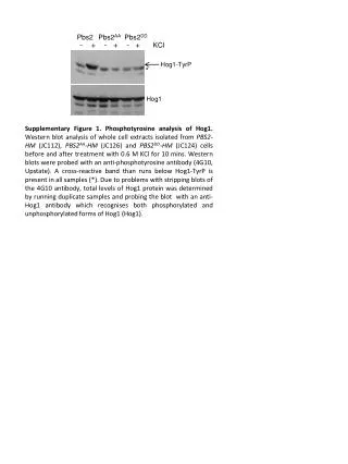
Phosphotyrosine Analysis of Hog1 in PBS2-HM, PBS2AA-HM, and PBS2DD-HM Cells Post KCl Treatment
This study investigates the phosphotyrosine levels of the Hog1 protein in whole cell extracts from PBS2-HM, PBS2AA-HM, and PBS2DD-HM cells. Cells were treated with 0.6 M KCl for 10 minutes, followed by Western blot analysis probing with an anti-phosphotyrosine antibody (4G10). Notably, a cross-reactive band appears in all samples beneath the Hog1-TyrP band. As a result of challenges in stripping the blots, total Hog1 protein levels were evaluated using duplicate samples and an anti-Hog1 antibody, which detects both phosphorylated and unphosphorylated Hog1.

Phosphotyrosine Analysis of Hog1 in PBS2-HM, PBS2AA-HM, and PBS2DD-HM Cells Post KCl Treatment
E N D
Presentation Transcript
Pbs2 Pbs2AA Pbs2DD - + - + - + KCl Hog1-TyrP * Hog1 Supplementary Figure 1. Phosphotyrosine analysis of Hog1. Western blot analysis of whole cell extracts isolated from PBS2-HM(JC112), PBS2AA-HM (JC126) andPBS2DD-HM(JC124) cells before and after treatment with 0.6 M KCl for 10 mins. Western blots were probed with an anti-phosphotyrosine antibody (4G10, Upstate). A cross-reactive band than runs below Hog1-TyrP is present in all samples (*). Due to problems with stripping blots of the 4G10 antibody, total levels of Hog1 protein was determined by running duplicate samples and probing the blot with an anti-Hog1 antibody which recognises both phosphorylated and unphosphorylated forms of Hog1 (Hog1).