Download
1 / 41
410 likes | 537 Views
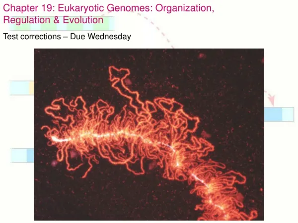
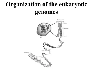
THE ORGANIZATION AND CONTROL OF EUKARYOTIC GENOMES and BIOTECHNOLOGY. Chapter 18 and 20. GENOME ORGANIZATION. 1.5% of DNA in humans codes for protein, tRNA, or rRNA 24% introns and regulatory proteins Most is repetitive DNA (59%) Unique noncoding is 15%.

E N D
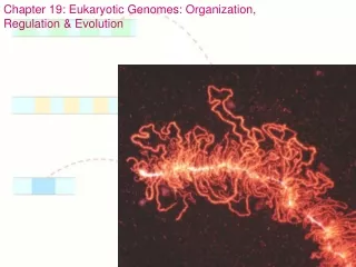
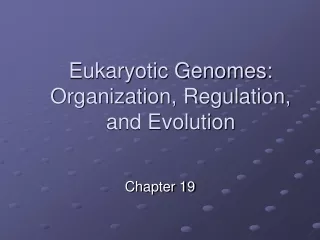
THE ORGANIZATION AND CONTROL OF EUKARYOTIC GENOMES and BIOTECHNOLOGY Chapter 18 and 20
GENOME ORGANIZATION • 1.5% of DNA in humans codes for protein, tRNA, or rRNA • 24% introns and regulatory proteins • Most is repetitive DNA (59%) • Unique noncoding is 15%
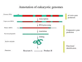
Figure 19.7 Opportunities for the control of gene expression in eukaryotic cells
Fig. 18-6a Signal NUCLEUS Chromatin Chromatin modification DNA Gene available for transcription Gene Transcription Exon RNA Primary transcript Intron RNA processing Tail mRNA in nucleus Cap Transport to cytoplasm CYTOPLASM
Fig. 18-6b CYTOPLASM mRNA in cytoplasm Translation Degradation of mRNA Polypeptide Protein processing Active protein Degradation of protein Transport to cellular destination Cellular function
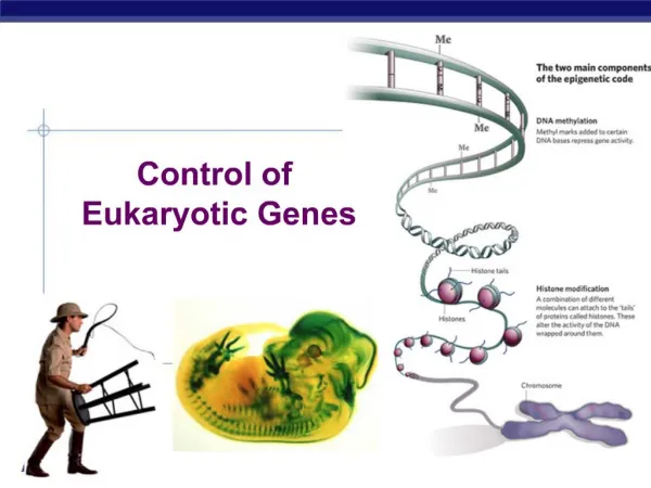
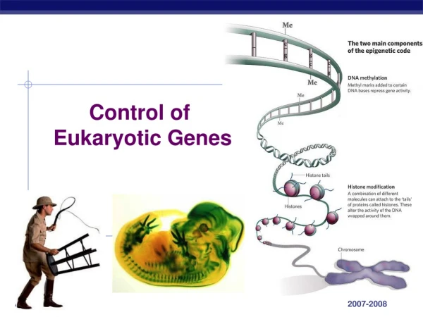
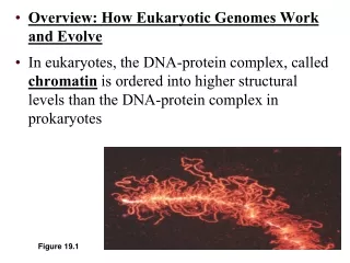
GENE Expression and REGULATION • Typical human cell expresses only 20% of its genes • Regulation of chromosome structure • Histone acetylation (-COCH3) loosens chromatin so transcription can occur • DNA methylation (-CH3) inactivates DNA • Responsible for X-inactivation • Genomic imprinting – in mammals, methylation turns off paternal or maternal allele of certain genes at start of development
Fig. 18-7 Histone tails Amino acids available for chemical modification DNA double helix (a) Histone tails protrude outward from a nucleosome Unacetylated histones Acetylated histones (b) Acetylation of histone tails promotes loose chromatin structure that permits transcription
Figure 19.7 Opportunities for the control of gene expression in eukaryotic cells
Regulation of transcription • Transcription factors – protein to protein recognition at TATA box needed for transcription to take place • Control elements– upstream of gene in promoter • Enhancers – far upstream and far downstream of gene; bind to transcription factors
Activator – transcription factors bound to enhancer that stimulate transcription • Not many different control elements so the combination of control elements regulates gene action • Different genes can be turned on by same activator
Fig. 18-9-3 Promoter Activators Gene DNA Distal control element Enhancer TATA box General transcription factors DNA-bending protein Group of mediator proteins RNA polymerase II RNA polymerase II Transcription initiation complex RNA synthesis
Post transcription regulation • RNA processing • Lifespan of mRNA in cell controls expression • Removal of caps leads to mRNA destruction • Translation prevented by protein not letting ribosome to attach to mRNA
Figure 19.7 Opportunities for the control of gene expression in eukaryotic cells
Biotechnology • Definition: The manipulation of organisms and their components to perform practical tasks or provide useful products • Used in areas from agriculture to criminal law • Most important in DNA Technology • Manipulation and analysis of DNA
Problems scientists have with studying DNA • DNA is huge and one strand usually carries many genes • Genes only occupy a small portion of DNA • DNA molecules are structurally and chemically homogenous • Need to develop methods for preparing well-defined gene-sized pieces of DNA with many copies • DNA cloning and the use of recombinant DNA is the answer
Using Restriction Enzymes to Make Recombinant DNA • Bacterial restriction enzymes cut DNA molecules at specific DNA sequences called restriction sites • A restriction enzyme usually makes many cuts, yielding restriction fragments • The most useful restriction enzymes cut DNA in a staggered way, producing fragments with “sticky ends” that bond with complementary sticky ends of other fragments • DNA ligaseis an enzyme that seals the bonds between restriction fragments
Fig. 20-3-3 Restriction site 5 3 3 5 DNA Restriction enzymecuts sugar-phosphatebackbones. 1 Sticky end DNA fragment addedfrom another moleculecut by same enzyme.Base pairing occurs. 2 One possible combination DNA ligaseseals strands. 3 Recombinant DNA molecule
Amplifying DNA in Vitro: The Polymerase Chain Reaction (PCR) • The polymerase chain reaction, PCR, can produce many copies of a specific target segment of DNA • A three-step cycle—heating, cooling, and replication—brings about a chain reaction that produces an exponentially growing population of identical DNA molecules
Fig. 20-8 3 5 TECHNIQUE Targetsequence 3 5 Genomic DNA 1 5 3 Denaturation 5 3 2 Annealing Cycle 1yields 2 molecules Primers 3 Extension Newnucleo-tides Cycle 2yields 4 molecules Cycle 3yields 8 molecules;2 molecules(in whiteboxes)match targetsequence
Gel Electrophoresis • One indirect method of rapidly analyzing and comparing genomes is gel electrophoresis • This technique uses a gel as a molecular sieve to separate nucleic acids or proteins by size • A current is applied that causes charged molecules to move through the gel • Molecules are sorted into “bands” by their size
Fig. 20-9 TECHNIQUE Powersource Mixture ofDNA mol-ecules ofdifferentsizes – Cathode Anode + Gel 1 Powersource – + Longermolecules 2 Shortermolecules RESULTS
Fig. 20-9a TECHNIQUE Powersource Mixture ofDNA mol-ecules ofdifferentsizes Anode Cathode – + Gel 1 Powersource – + Longermolecules 2 Shortermolecules
Gel Electrophoresis • In restriction fragment analysis, DNA fragments produced by restriction enzymes digestion of a DNA molecule are sorted by gel electrophoresis • Restriction fragment analysis is useful for comparing two different DNA molecules, such as two alleles for a gene • The procedure is also used to prepare pure samples of individual fragments
Fig. 20-10 Normal -globin allele Normalallele Sickle-cellallele 175 bp Large fragment 201 bp DdeI DdeI DdeI DdeI Largefragment Sickle-cell mutant -globin allele 376 bp 201 bp175 bp Large fragment 376 bp DdeI DdeI DdeI (a) DdeI restriction sites in normal and sickle-cell alleles of -globin gene (b) Electrophoresis of restriction fragments from normal and sickle-cell alleles
Studying the Expression of Interacting Groups of Genes • Automation has allowed scientists to measure expression of thousands of genes at one time using DNA microarray assays • DNA microarray assays compare patterns of gene expression in different tissues, at different times, or under different conditions
Fig. 20-15 TECHNIQUE Tissue sample 1 Isolate mRNA. 2 Make cDNA by reversetranscription, usingfluorescently labelednucleotides. mRNA molecules Labeled cDNA molecules(single strands) DNA fragmentsrepresentingspecific genes 3 Apply the cDNA mixture to amicroarray, a different gene ineach spot. The cDNA hybridizeswith any complementary DNA onthe microarray. DNA microarray DNA microarraywith 2,400human genes 4 Rinse off excess cDNA; scanmicroarray for fluorescence.Each fluorescent spot represents agene expressed in the tissue sample.
Diagnosis of Diseases • Scientists can diagnose many human genetic disorders by using PCR and primers corresponding to cloned disease genes, then sequencing the amplified product to look for the disease-causing mutation • Genetic disorders can also be tested for using genetic markers that are linked to the disease-causing allele
Single nucleotide polymorphisms (SNPs) are useful genetic markers • These are single base-pair sites that vary in a population • When a restriction enzyme is added, SNPs result in DNA fragments with different lengths, or restriction fragment length polymorphism (RFLP)
Fig. 20-21 DNA T Normal allele SNP C Disease-causingallele
Concept 20.1: DNA cloning yields multiple copies of a gene or other DNA segment • To work directly with specific genes, scientists prepare gene-sized pieces of DNA in identical copies, a process called DNA cloning • Most methods for cloning pieces of DNA in the laboratory share general features, such as the use of bacteria and their plasmids • Plasmids are small circular DNA molecules that replicate separately from the bacterial chromosome • Cloned genes are useful for making copies of a particular gene and producing a protein product
Cell containing geneof interest Bacterium 1 Gene inserted intoplasmid Bacterialchromosome Plasmid Gene ofinterest RecombinantDNA (plasmid) DNA of chromosome 2 Plasmid put intobacterial cell Recombinantbacterium DNA Cloning 3 Host cell grown in cultureto form a clone of cellscontaining the “cloned”gene of interest Gene ofInterest Protein expressedby gene of interest Copies of gene Protein harvested Basic research andvarious applications 4 Basicresearchon protein Basicresearchon gene Gene used to alter bacteria for cleaning up toxic waste Gene for pest resistance inserted into plants Protein dissolvesblood clots in heartattack therapy Human growth hor-mone treats stuntedgrowth
Problems with cloning eukaryotic genes • There are differences in gene expression between prokaryotes and eukaryotes • Ex. Prokaryotes have no machinery to cut out introns • We use cDNA to overcome this problem • We can also use yeast and fungus to clone although there are still problems associated with these two processes as well.
Storing Cloned Genes in DNA Libraries • A genomic library that is made using bacteria is the collection of recombinant vector clones produced by cloning DNA fragments from an entire genome • A genomic library that is made using bacteriophages is stored as a collection of phage clones
Fig. 20-5 Foreign genomecut up withrestrictionenzyme Large insertwith many genes Large plasmid or BACclone Recombinantphage DNA Bacterial clones Recombinantplasmids Phageclones (a) Plasmid library (b) Phage library (c) A library of bacterial artificial chromosome (BAC) clones
Storing Cloned Genes in DNA Libraries • A complementary DNA (cDNA) library is made by cloning DNA made in vitro by reverse transcription of all the mRNA produced by a particular cell • A cDNA library represents only part of the genome—only the subset of genes transcribed into mRNA in the original cells • Screening a Library for Clones Carrying a Gene of Interest using nucleic acid hybridization
Fig. 20-6-5 DNA innucleus mRNAs in cytoplasm Reversetranscriptase Poly-A tail mRNA Primer DNAstrand DegradedmRNA DNA polymerase cDNA
A probe can be synthesized that is complementary to the gene of interest • For example, if the desired gene is – Then we would synthesize this probe … … 5 G G C T A A C T T A G C 3 C C G A T T G A A T C G 5 3
Fig. 20-7 • TECHNIQUE Radioactivelylabeled probemolecules ProbeDNA Gene ofinterest Multiwell platesholding library clones Single-strandedDNA from cell Film Nylon membrane Nylonmembrane Location ofDNA with thecomplementarysequence
Human Gene Therapy • Gene therapy is the alteration of an afflicted individual’s genes • Gene therapy holds great potential for treating disorders traceable to a single defective gene • Vectors are used for delivery of genes into specific types of cells, for example bone marrow • Gene therapy raises ethical questions, such as whether human germ-line cells should be treated to correct the defect in future generations
Fig. 20-22 Clonedgene Insert RNA version of normal alleleinto retrovirus. 1 Viral RNA Let retrovirus infect bone marrow cellsthat have been removed from thepatient and cultured. 2 Retroviruscapsid Viral DNA carrying the normalallele inserts into chromosome. 3 Bonemarrowcell frompatient Bonemarrow Inject engineeredcells into patient. 4