Download
1 / 43
480 likes | 1.89k Views
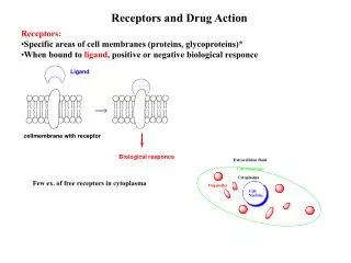
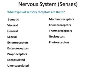
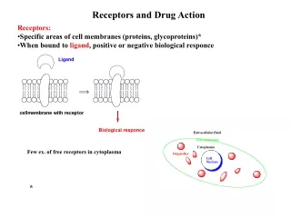
Types of drug receptors Practically all receptors are proteins : Enzymes Ion channels Ligand-gated channels: Ion channels that open upon binding of a mediator Voltage-gated channels: Ion channels that are not normally controlled by ligand binding but by changes in the membrane potential

E N D
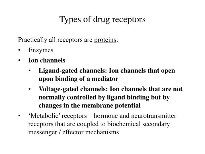
Types of drug receptors • Practically all receptors are proteins: • Enzymes • Ion channels • Ligand-gated channels: Ion channels that open upon binding of a mediator • Voltage-gated channels: Ion channels that are not normally controlled by ligand binding but by changes in the membrane potential • ‘Metabolic’ receptors – hormone and neurotransmitter receptors that are coupled to biochemical secondary messenger / effector mechanisms
Physiology and pharmacology of membrane excitation • Excitable cell types: • Nerve cells • Myelinated nerve fibers (fast transmission) • Non-myelinated nerve fibers (slow transmission) • Muscle cells • Skeletal muscle • Heart muscle • Smooth muscle “striated”
Membrane potentials and excitability • Both excitable and non-excitable cell membranes have an electrical potential across their cytoplasmic membranes • The membrane potential chiefly depends on the asymmetric distribution of sodium and potassium ions, and with some cells calcium ions across the cell membrane • In the ‘ground state’, the orientation of the membrane potential is negative inside
3 Na+ Na+ Ca++ Glucose How is the asymmetric distribution of ions across the membrane maintained? 2 K+ ADP + Pi ATP 3 Na+ K+ Cl- K+ Na+
Ionic basis of membrane potentials and excitability • In the resting state of excitable cells – and throughout in the non-excitable cells – the interior of the cell is electrically negative against the outside • Electrical excitation (the ‘action potential’) consists in a brief, transient reversal of the orientation of the membrane potential • Both the resting potential and the action potential are diffusion potentials
- - - - - - - - - - + + + + + + + + + + Diffusion potentials (1) no potential (electroneutrality)
- - - - - - - - - - + + + + + + + + + + Diffusion potentials (2) still no potential (electroneutrality) + -
- + Diffusion potentials (3) negative positive - + - + - + - + - + - + - + - + - +
- - + + - - + + Diffusion potentials (4) Driving force 1: Entropy (equalize concentrations on both sides) Driving force 2: Electroneutrality (equalize charges on both sides)
Cout ln E = Cin E: R: T: F: z: ln: Cin, Cout The equilibrium diffusion potential Gas constant (8.31 J K-1mol-1) Absolute temperature (K) Faraday constant (96500 Coulomb/mole) Number of charges of single ion (1 with K+ and Na+, 2 with Ca++, -1 with Cl-) Natural logarithm (base: e = 2.71828) Inside and outside concentrations of the diffusible ion species R T z F The Nernst equation describes the diffusion potential at equilibrium
Inside Outside Equilibrium potential Na+15 mM 150 mM +60 mV K+ 150 mM 6 mM -90 mV What if there are multiple diffusible ions? (1) Intra- and extracellular cation concentrations: Actual resting membrane potential: -70 mV
Cout ln Nernst equation: E = Cin R T z F PK [K+]out + PNa [Na+]out R T ln E = PK [K+]in + PNa [Na+]in F What if there are multiple diffusible ions? (2) Goldman equation (special case for Na and K):
PK [K+]out + PNa [Na+]out R T ln E = PK [K+]in + PNa [Na+]in F The Goldman equation and the role of ion channels P = Permeability – this is where the ion channels come in
PK [K+]out + PNa [Na+]out R T ln E = PK [K+]in + PNa [Na+]in F The Goldman equation and the role of ion channels (2) change don’t change
+ + - K+ - Na+ - K+ Na+ - - K+ Na+ - - K+ Na+ - - Na+ - The cellular resting potential is essentially a potassium potential negative positive K+
+ + Voltage-gated sodium channels will open upon reversal of the resting membrane potential negative positive Na+ negative positive
+ + + Voltage-gated sodium channels propagate the action potential negative positive - - - - Na+ Na+ outside inside Na+ K+ K+ K+ K+ - - - - positive negative spreading action potential
- 55 mV Firing level - 70 mV - 85 mV Electrical depolarization of nerve fibers can trigger action potentials External stimuli of varying amplitude time (ms)
ENa (+60 mV) Repolarization: K channels open Na+ channels close Depolarization: Na channels open (PNa > PK) Hyperpolarization: Na channels closed (PK >> PNa) Resting potential: PK > PNa EK (-90 mV) The Goldman equation and the action potential
Set voltage externally Measure resulting current across channel Planar lipid membranes allow observation of individual channels
Multiple opening events of a single channel in a planar lipid bilayer Externally applied voltage Multiple, successive observations open state base line / closed averaged trace Current Time
Patch clamping pipette channel cell
Cell attached mode seal
Whole cell mode suction seal
Excised patch mode seal cell ripped apart
Questions: • How is the action potential initiated ? • How is the action potential terminated ?
Action potential: Termination (1) • The ion flux through the voltage-gated Na+ channel is countered by a voltage-gated K+ channel that responds more slowly to depolarization • Both channels spontaneously inactivate Resulting membrane potential Na+ influx K+ efflux duration: a few milliseconds
Depolarization Spontaneous inactivation Closed Open Inactivated Slow reactivation after membrane repolarization Action potential: Termination (2) Voltage-gated channels cycle between 3 distinguishable functional states
Structural model of a Kv channel Extracellular space Cytosol
+ + + - - - + + + + + + + + + + + + + + + + + + + + - - - + + + K+ K+ The KV channel’s opening gate is located in the membrane
+ + + + + + + + + + + + + + + + - - - - - - + + The KV channel in the resting state
The KV channel in the open state - - - + + + + + + + + + + + + + + +
- - - + + + + + + + + + + + + + + + The KV channel in the inactivated state
Action potential: Initiation • In a resting cell, an action potential can be initiated in a variety of ways: • By synaptic transmission. Examples: Signal conduction from one nerve cell to another, from nerve cell to muscle cell • By spontaneous, rhythmic membrane depolarization. Example: Specialized cells in heart and smooth muscle • By electrical coupling to a neighboring cell via gap junctions. Example: Heart muscle, smooth muscle
+ Na+ K+ presynaptic action potential Synaptic excitation ENa Firing level EK Presynaptic terminal synaptic cleft Postsynaptic terminal
- - + Na+ + Synaptic excitation (2) - Na+ Na+ In synapses, ligand-gated channels open upon binding of neurotransmitters and initiate the action potential in the post-synaptic membrane
+ + Action potential initiation in heart pacemaker cells (negative charge left behind) Ca++ Ca++ K+ Ca++ K+ K+ In heart pacemaker cells, two types of calcium channels lead to spontaneous depolarization
Action potential initiation in heart pacemaker cells K+ 0 mV Ca++L -40 mV Ca++T -60 mV slow, spontaneous prepotential
Cell excitation by electrical couplingacross gap junctions - - - - - + + + + - - - - - + + + + Gap junction
negative positive - + + - - + + - - + - + Permeating anions leave behind excess positive charge Permeating cations leave behind excess negative charge negative positive R * T PK* CK,out + PNa* CNa,out + PCl, * CCl, in F * ln E = PK* CK,in + PNa* CNa,in + PCl, * CCl, out What about anions? Opposite charge affects the Goldman equation:
Inside cell Outside cell Equilibrium potential Na+15 mM 150 mM +60 mV K+ 150 mM 6 mM -90 mV Cl- 9 mM 125 mM -70 mV Ca++ 100 nM 1.3 mM +130 mV Intra- and extracellular ion concentrations • Opening of sodium or calcium channels will increase the membrane potential (depolarization) • Opening of potassium or chloride channels will lower the membrane potential (repolarization or hyperpolarization)
+ Cl- Sodium and chloride in excitatory and inhibitory synapses Na+ positive negative