Download
1 / 37
370 likes | 724 Views
Shoulder and Upper Arm Pathologies. Chapter 16. Clinical Anatomy. Bony anatomy Manubrium Jugular notch Clavicular notch Clavicle Scapula Subscapular fossa Vertebral border Inferior and superior angle Scapular spine Supraspinous fossa Acromion process Coracoid process.

E N D
Shoulder and Upper Arm Pathologies Chapter 16
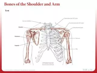
Clinical Anatomy Bony anatomy Manubrium Jugular notch Clavicular notch Clavicle Scapula Subscapular fossa Vertebral border Inferior and superior angle Scapular spine Supraspinous fossa Acromion process Coracoid process
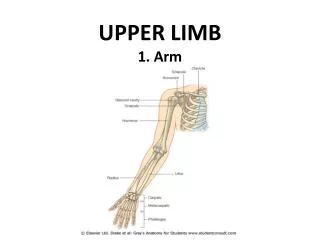
Clinical Anatomy Bony anatomy Humerus Humeral head Bicipital groove Greater tuberosity Lesser tuberosity Surgical neck Deltoid tuberosity
Clinical Anatomy Joints of the shoulder complex Glenohumeral joint (GH) Acromioclavicular joint (AC) Sternoclavicular joint (SC) Scapulothoracic articulation
Clinical Anatomy Bursa of the shoulder complex Subacromial bursa Above supraspinatus tendon Buffers tendons contact with acromion process and the coracoacromial ligament Inflammed bursa can lead to RTC impingement Subdeltoid bursa
Clinical Examination of Shoulder Injuries Past medical history Previous history AC or GH injury can alter biomechanics Cervical spine pathology Can radiate pain to upper extremity History of the present condition Location of the pain Onset Activity and injury mechanism Symptoms
Clinical Examination of Shoulder Injuries Inspection Functional assessment Pain in follow-through Pain in cocked position Pain in deceleration Loss of control and/or velocity
Clinical Examination of Shoulder Injuries Inspection Anterior shoulders Level of the shoulders Position of the head Position of the arm Contour of the clavicles Symmetry of the deltoid muscle group Anterior humerus and biceps brachii muscle group
Clinical Examination of Shoulder Injuries Inspection Posterior structures Alignment of the vertebral column Position of the scapula Sprengel’s deformity — congenitally undescended scapula Muscle development Position of the humerus
Clinical Examination of Shoulder Injuries Palpation of the anterior shoulder Jugular notch Sternoclavicular joint Clavicular shaft Acromion process and AC joint Coracoid process Humeral head Greater tuberosity Lesser tuberosity Bicipital grove Humeral shaft Pectoralis major Pectoralis minor Coracobrachialis Deltoid group Biceps brachii Long head of the biceps Short head of the biceps
Clinical Examination of Shoulder Injuries Palpation of the posterior shoulder Spine of the scapula Superior angle Inferior angle Infraspinatus Teres minor Supraspinatus Teres major Rhomboid major Rhomboid minor Levator scapulae Trapezius Latissimus dorsi Posterior deltoid Triceps brachii
Clinical Examination of Shoulder Injuries Joint and muscle function assessment Active range of motion (AROM) Flexion and extension Abduction and adduction Internal and external rotation Horizontal adduction and abduction Manual muscle testing (MMT) Scapular movements Passive range of motion (PROM) Same motions as AROM
Clinical Examination of Shoulder Injuries Joint stability tests Sternoclavicular joint play Test for acromioclavicular joint laxity Test for glenohumeral joint laxity Neurologic testing Upper quarter screen Referred pain from visceral organs
Acromioclavicular joint pathology “Separated shoulder” MOI: FOOSH, blow to superior acromion process Classification of sprains depends on structures involved, degree of instability, and direction of displaced clavicle Pathologies of the Shoulder and Related Special Tests
Glenohumeral instability Anterior instability Posterior instability Inferior instability Multidirectional instability Pathologies of the Shoulder and Related Special Tests
Rotator cuff pathology Impingement syndrome Rotator cuff tendinopathy Subacromial bursitis Pathologies of the Shoulder and Related Special Tests
Biceps tendon pathology Bicipital tendinopathy Causes RTC dysfunction Impingement Superior labrum anterior to posterior lesions (SLAP lesions) Tears of the superior aspect of the glenoid labrum that extend anteriorly and posteriorly to the biceps insertion Pathologies of the Shoulder and Related Special Tests
On-Field Examination of Shoulder Injuries On-field history Location of pain Upper shoulder AC sprain Trapezius Brachial plexus injury MOI Internal or external rotation (with abduction) GH joint discloation or subluxation FOOSH Clavicular fracture, AC sprain, SC sprain On-field inspection Arm posture Arm splinted against torso Arm hanging limply at the side Arm “locked” Gross deformity
On-Field Examination of Shoulder Injuries On-field palpation Position of the humeral head AC joint alignment Clavicle Sternoclavicular joint Humerus Additional on-field tests If joint dislocation or bony fracture have been ruled out Apley’s scratch test (see Box 13–3) can be used as a gross assessment of the athlete’s willingness to move the involved extremity and the amount of motion
Scapular fracture Body of the scapula Glenoid fossa Glenoid neck Coracoid process Management Immobilize the arm on the affected side in a comfortable position Athlete then is transported GH dislocation also needs a radiographic evaluation to rule out a secondary fracture to the glenoid or coracoid process Initial Management of On-Field Shoulder Injuries
Clavicular fracture Immobilization using a sling or triangular bandage Transport for definitive diagnosis Sternoclavicular joint injuries Neurologic and vascular examination of the extremity and carotid artery Involved arm is immobilized Athlete is immediately transported to an emergency medical facility Acromioclavicular joint injuries Immobilize in a position that lessens the displacement between the clavicle and the acromial process Protect joint with additional padding during activity On-Field Examination of Shoulder Injuries
Glenohumeral dislocations • Monitor the distal pulses, check for circulation in the fingertips, and perform a sensory screen • Arm is fixed in the position it has assumed • Reductions of GH dislocations should only be performed by those who are trained to do so • Forced reduction of the humeral head may damage the glenoid fossa, the coracoid process, or the neurovascular structures in the area. Following reduction, assess distal pulse and active range of motion, avoiding external rotation and abduction. Stabilize the shoulder using a sling, and refer the athlete for further examination. • Humeral fractures • Splint in position found using moldable splint or vacuum splint • Leave wrist and fingers exposed to check circulation • Transport