Overview of Systemic Mycoses: Histoplasmosis
Learn about histoplasmosis, a systemic mycosis caused by Histoplasma capsulatum. Explore its etiology, pathogenesis, clinical types, major forms, diagnosis methods, and more.

Overview of Systemic Mycoses: Histoplasmosis
E N D
Presentation Transcript
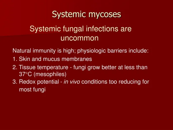
Causative agents: thermally dimorphic fungi that exist in nature, soil. Geographic distribution varies. Inhalation pulmonary inf. dissemination No evidence of transmission among humans or animals. Otherwise healthy individuals are infected. SYSTEMIC MYCOSES General features
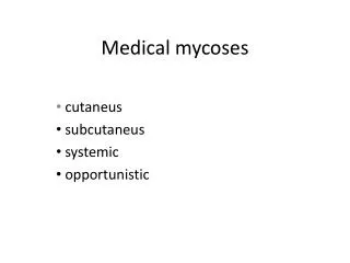
Histoplasmosis Blastomycosis Coccidioidomycosis Paracoccidioidomycosis
Histoplasmosis • An intracellular mycotic infection of the reticuloendothelial system. • Distribution: World-wide, especially U.S.A. Sporadic cases do occur in Australia. • Histoplasmosis is a significant occupational disease in bat caves in Mexico when workers harvest the guano for fertilizer. • In the endemic area the majority of patients who develop histoplasmosis (95%) are asymptomatic.
Aetiological agent: Histoplasma capsulatum • Member of the phylum Ascomycota. • Naturally found in fecal-contaminated soils- especially from soil enriched with excreta from chicken, starlings and bats. • Birds and bats appear to be reservoirs.
Dimorphic fungus • Sexual multi-cellular saprophytic mycelia • Asexual single-celled parasitic yeast • Mycelial form is most commonly found in the environment.
Form asexual macro- and microconidia • Also borne by hyphae in the mycelial form • Yeast cells have white, thin-walled, oval bodies
Pathogenesis • Infection begins with inhalation of microconidia or hyphal fragments • Mycelial form transforms into yeast form • Triggered by elevated temperatures and increased cysteine levels Pathogenesis
Yeast cells are phagocytized by host immune system • M. capsulatum is able to survive phagocytosis • Calcium-binding protein, a cytoplasmic enzyme, a peroxisomal enzyme, and immunogenic M antigen are involved • Apoptosis of infected macrophages allow H. capsulatum to spread.
Clinical Types; • Histoplasmosis is a systemic disease, mostly of the reticulo-endothelial system, manifesting itself in the bone marrow, lungs, liver, and the spleen. • Hepatosplenomegaly is the primary sign in children, while in adults, histoplasmosis more commonly appears as pulmonary disease. • There is generally complete recovery from the acute pulmonary form (another "flu-like" illness).
However, if untreated, it disseminate. The disseminated form of disease is usually fatal. • Patients will first notice shortness of breath and a cough which becomes productive. • The sputum may be purulent or bloody. • Patients will become anorexic and lose weight. They have night sweats. ….sounds like tuberculosis.
Major forms of histoplasmosis; • Pulmonary and disseminated • Pulmonary histoplasmosis occurs when microconidia or mycelial fragments are inhaled • Form lesions in the hilar and/or mediastinal nodes • Many types of pulmonary histoplasmosis; • Asymptomatic pulmonary histoplasmosis • Acute pulmonary histoplasmosis • Mediastinal granuloma • Fibrosing mediastinitis • Chronic cavitary pulmonary histoplasmosis
Asymptomatic pulmnary histoplasmosis • 99% of infected people display no symptoms • May have mild “illness” • Diagnosed using radiography, CT scans, or biopsies • Acute pulmonary histoplasmosis • Higher level exposure to H. capsulatum • Patients display fever, malaise, headache, dyspnea, and other respiratory problems
Mediastinal granuloma • Substantial enlargement of a large number of mediastinal lymph nodes • Can impede airways or the superior vena cava • Often matted together and necrotic • Patients have severe chest pain when breathing • Fibrosing mediastinitis • Uncontrolled immune response to necrotizing nodes causes fibrosis around mediastinal lymph nodes • Patients display worsening dyspnea, cough, hemoptysis, and chest pain • Superior vena cava obstruction and heart failure can occur
Chronic pulmonary histoplasmosis • Exclusive to older patients with emphysema • H. capsulatum infection near emphysematous bullae form a cavity • The cavity progressively grows and spreads from lobe to lobe to form more cavities • Patients display fatigue, fever, anorexia, weight loss, hemoptysis, and dyspnea
Disseminated Histoplasmosis • Occurs primarily in immunocompromised individuals • In healthy individuals, H. capsulatum is similar to tuberculosis • In immunocompromised individuals, it is able to spread from the lungs into other organs • Patients display fever, malaise, and occasionally petechiae or skin lesions (cutaneous histoplasmosis) • Tests often reveal mucous membrane ulcerations, simultaneous enlargement of the liver and spleen, and enlarged lymph nodes
Histoplasmosis of the lower gum showing ulcer around base of tooth
Laboratory diagnosis • Clinical specimens; depend on the presentation of the disease • Sputum or Bronchial alveolar lavage, if it is pulmonary disease • Biopsy material from the affected organ. • Bone marrow • Blood • Methods: • Microscopy • Culture • Serology
Microscopy • The yeast is usually found in monocytes or in PMNL's. • In peripheral blood, H. capsulatum appears as a small yeast about 5-6 µ in diameter.
Tissue section stained by Grocott's methenamine silver stain
Culture • Media used: Sabouraud’s dextrose agar & BHI blood agar. • At 37ºC: Yeast phase . • It is a white to tan colony. • The yeast cell is 5-6 µ in diameter and slightly oval. • At 25ºC: The mold phase characterized by thin, branching, septate hyphae that produce microconidia and a very distinct spore called a tuberculate macroconidium. • To confirm the diagnosis, the organism must be converted from yeast to mycelium or vice-versa or by using DNA probe
Culture of Histoplasma capsulatum on Sabouraud's dextrose agar showing a white suede-like colony
Culture of Histoplasma capsulatum on Sabouraud's dextrose agar showing a white suede-like colony with a pale yellow-brown reverse.
Microscopic morphology of the saprophytic or mycelial form of Histoplasma capsulatum showing characteristic large, rounded, single-celled, tuberculate macroconidia
Serological Test • Serology for histoplasmosis is a little more complicated than for other mycoses, There are 4 tests: • Latex agglutination • Complement Fixation • Immunodiffusion • EIA
Treatment; • The drug of choice is amphotericin B • Itraconazole and Voriconazole is now also being used