
Download
1 / 1
Download Presentation 
An Image/Link below is provided (as is) to download presentation
Download Policy: Content on the Website is provided to you AS IS for your information and personal use and may not be sold / licensed / shared on other websites without getting consent from its author.
Content is provided to you AS IS for your information and personal use only.
Download presentation by click this link.
While downloading, if for some reason you are not able to download a presentation, the publisher may have deleted the file from their server.
During download, if you can't get a presentation, the file might be deleted by the publisher.
E N D
Presentation Transcript
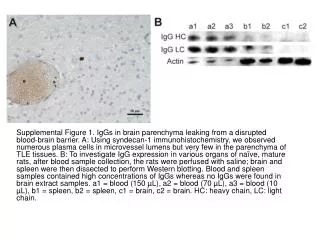
Supplemental Figure 1. IgGs in brain parenchyma leaking from a disrupted blood-brain barrier. A: Using syndecan-1 immunohistochemistry, we observed numerous plasma cells in microvessel lumens but very few in the parenchyma of TLE tissues. B: To investigate IgG expression in various organs of naïve, mature rats, after blood sample collection, the rats were perfused with saline; brain and spleen were then dissected to perform Western blotting. Blood and spleen samples contained high concentrations of IgGs whereas no IgGs were found in brain extract samples. a1 = blood (150 µL), a2 = blood (70 µL), a3 = blood (10 µL), b1 = spleen, b2 = spleen, c1 = brain, c2 = brain. HC: heavy chain, LC: light chain.
More Related