1 / 1

CPT-induced Apoptosis in LLCPK-1a Cells Revealed by Fluorescence Microscopy

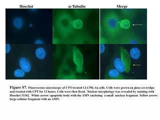
This figure shows the effects of CPT treatment on LLCPK-1a cells, demonstrating nuclear morphology using Hoechst 33342 staining. It highlights apoptotic bodies and cellular fragments as indicated by white and yellow arrows, respectively.

Download Presentation

CPT-induced Apoptosis in LLCPK-1a Cells Revealed by Fluorescence Microscopy

An Image/Link below is provided (as is) to download presentation Download Policy: Content on the Website is provided to you AS IS for your information and personal use and may not be sold / licensed / shared on other websites without getting consent from its author. Content is provided to you AS IS for your information and personal use only. Download presentation by click this link. While downloading, if for some reason you are not able to download a presentation, the publisher may have deleted the file from their server. During download, if you can't get a presentation, the file might be deleted by the publisher.

E N D

Presentation Transcript


  1. Hoechst a-Tubulin Merge Figure S7. Fluorescence microscopy of CPT-treated LLCPK-1a cells. Cells were grown on glass coverslips and treated with CPT for 12 hours. Cells were then fixed. Nuclear morphology was revealed by staining with Hoechst 33342. White arrow: apoptotic body with the AMN enclosing a small nuclear fragment. Yellow arrow: large cellular fragment with an AMN.

More Related