Download
1 / 52
600 likes | 1.01k Views
Kidney Stones: An Overview. Gerald Da Roza MD, MHSc, FRCPC March 15, 2010. Overview. Case Diagnosis of kidney stones Acute management Epidemiology Risk factors Work up and treatment Diet and kidney stones. Case – A Few Years Ago. 30 year old nephrology fellow

E N D
Kidney Stones: An Overview Gerald Da Roza MD, MHSc, FRCPC March 15, 2010
Overview • Case • Diagnosis of kidney stones • Acute management • Epidemiology • Risk factors • Work up and treatment • Diet and kidney stones
Case – A Few Years Ago • 30 year old nephrology fellow • Bright, hardworking, driven • Atrocious diet (hospital cafeteria and vending machines, no fruit and vegetables, ++ salt) • Drinks very little during daytime • Presents with acute onset of R costovertebral pain, radiating around to anterior abdomen, 10/10 in severity, nauseau and vomiting
Case – A Few Years Ago • Physical Exam • Tachycardia, normotensive, afebrile • ++ CVA and RUQ tenderness • Nil else • Investigations • U/A shows hematuria, • CBC, lytes urea, Cr normal
Diagnosis??? • Kidney Stone - Why? • DDx • Renal Cell Ca w/ blood clot • Renal Cyst w/ clot • Pyelonephritis • AAA/dissection • Ectopic Pregnancy (if female) • Intestinal Obstruction • Appendicitis
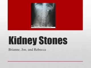
How do we make the diagnosis? • Investigative Options: • CT Scan • US • Abdominal Plain Film • MRI • IVP
Non-contrast Helical CT Scan • Gold standard • Sensitivity 95 %, Specificity 98% • Dual energy CT (DECT) is new imaging modality may be able to predict stone composition (future tx) • Helps determine if obstruction present • Provides alternate diagnosis in many cases • 33 percent had an alternate diagnosis not suspected on clinical grounds, one-half of whom had significant disease • Only misses stones due to protease inhibitors
Ultrasound • Procedure of choice for pts who should avoid radiation • pregnant women and possibly women of childbearing age • Sensitive for the diagnosis of obstruction • Can detect radiolucent stones missed on x-ray • May miss small stones and ureteral stones
Abdominal X-ray • will identify sufficiently large radiopaque stones • calcium, struvite, and cystine stones • will miss radiolucent uric acid stones • may miss small stones or stones overlying bony structures • will not detect obstruction
Other • Intravenous Pyelogram (IVP) • higher sensitivity and specificity than plain film for the • provides data about the degree of obstruction • previously the diagnostic procedure of choice, no longer because of potential contrast rxn, lower sens, higher radiation • Magnetic resonance imaging • rarely used during the management of stone disease, except in the evaluation of pregnant patients, because this modality is not optimal for identifying stones.
Acute Management • Many pts with acute renal colic can be managed conservatively with pain medication (NSAIDs & Opiods) and hydration until the stone passes • If able to take oral medications and fluids can manage at home • Hospitalization required for those who cannot tolerate oral intake or who have uncontrollable pain or fever
Acute Management • Pts instructed to strain their urine for several days and bring in any stone that passes for analysis • will enable clinician to better plan preventive therapy • Data suggests faster stone passage tamsulosin • CCB is other option • Pts are re-imaged if spontaneous passage has not occurred.
Acute Management • Urgent urologic consultation warranted in: • Urosepsis • Acute renal failure • Anuria • Unyielding pain, nausea, or vomiting
Acute Management • Stone size major determinant of the likelihood of spontaneous stone passage, although stone location is also important • Most stones ≤4 mm in diameter pass spontaneously. For stones larger than 4 mm in diameter, there is a progressive decrease in the spontaneous passage rate, which is unlikely with stones ≥10 mm in diameter • Proximal ureteral stones are also less likely to pass spontaneously.
Acute Management • Referral to urology for potential intervention • stones larger than 10 mm in diameter • significant discomfort • significant obstruction or who have not passed the stone after four to six weeks
Urologic Options • Shock wave lithotripsy (SWL) • tx choice in 75% pts • works best for stones in renal pelvis and upper ureter • Ureteroscopic lithotripsy with electrohydraulic or laser probes • higher stone-free rates, but with an increased incidence of complications over shock wave lithotripsy • Percutaneous nephrolithotomy • Laparoscopic stone removal • Rarely needed
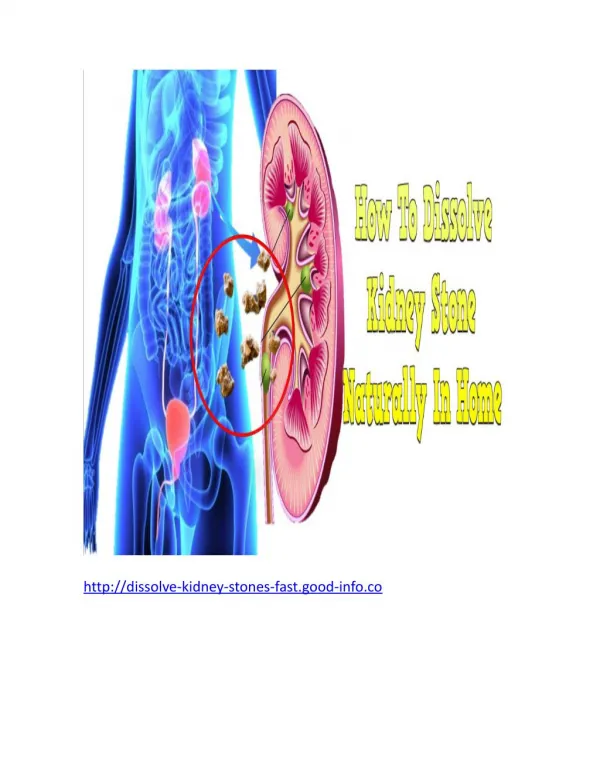
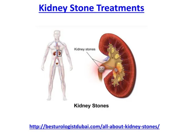
Kidney Stones - Epidemiology • Renal stones (nephrolithiasis) are a relatively common problem • In US, up to 12% of men and 5% of women will have at least one symptomatic stone by the age of 70
Clinical Presentations • Classic Sx • Renal Colic • Hematuria (gross or microscopic in majority if symptoms but not all) • Atypical Sx • Vague abdominal pain, nausea, urinary urgency or frequency, difficulty urinating, penile pain, or testicular pain. • Asymptomatic
Renal Colic • Varies from a mild and barely noticeable ache to discomfort that is so intense that requires parenteral analgesics • typically waxes and wanes in severity, and develops in waves or paroxysms that are related to movement of the stone in the ureter and associated ureteral spasm. • Paroxysms of severe pain usually last 20 to 60 minutes • Pain is thought to occur primarily from urinary obstruction with distention of the renal capsule.
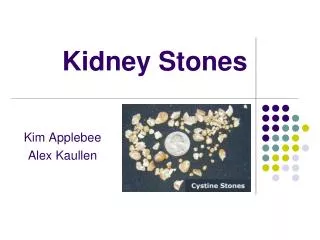
Stone Composition • 80% are Calcium Stones • Calcium Oxalate (majority) • Calcium Phosphate (Hydroxapetite stones)
Stone Composition • Uric acid • Struvite (magnesium ammonium phosphate) • only form in pts with chronic upper UTI d/t urease-producing organism: Proteus or Klebsiella • Cystine stones • only develop in pts with cystinuria (an AR disorder) due to the poor solubility of cystine in the urine • Mixed stone (eg, calcium oxalate and uric acid) • Other: indinavir, sulfadiazine, triamterene, acyclovir stone
Risk Factors for Stones • Historical • Anatomic • Dietary • Urinary
Historical Risk Factors • Prior History of Kidney Stones • 50% recurrence in 10 yrs • Family History of kidney stones • Twofold increase by Health professionals study • Individuals with enhanced enteric oxalate absorption • gastric bypass procedures, bariatric surgery, short bowel syndrome • Frequent upper urinary tract infections • Excessive physical exertion
Historical RF • Medical conditions assoc w/ stones: • Primary Hyperparathyroidism, Sarcoidosis • Gout, Obesity, DM (concentrated acidic urine) • HTN • RTA • Use of medications that may crystallize urine • Indinavir, acyclovir, sulfadiazine, triamterene
Anatomic RF • Medullary sponge kidney • Horseshoe kidney
Dietary Risk Factors • ? Low or High ? • Calcium • Fluids • Oxalate • Protein • Salt • Sucrose
Dietary Risk Factors • Low Calcium Intake • increases absorption & excretion of oxalate d/t less complexing with calcium in the intestinal lumen • Low fluid intake • Higher concentration of lithogenic factors in urine • Low potassium • Low phytate
Dietary Risk Factors • High oxalate intake • High animal protein intake • leads to hypercalciuria, hyperuricosuria, hypocitraturia, and inc urinary acid excretion • High sodium intake • High sucrose intake • may increase calcium and/or oxalate excretion • High Vitamin C Intake
Urinary Risk Factors • Low volume • Hypercalcuria • Hyperoxaluria • Hypocitraturia • Extremes of pH • pH greater than 7.5 is compatible with infection pH less than 5.5 favours uric acid lithiasis. • Urine culture +ve urease-producing organism (struvite) • Proteus or Klebsiella
Work Up & Treatment • Controversial whether evaluation and therapy warranted or cost effective after the first stone or only in patients with: • Active stone disease • formation of new stones, increase in size of old stones, or the continued passage of gravel • Multiple stones at first presentation • Pts with a strong family history of stones
Approaches • Limited Evaluation • Targeted Evaluation • base the extent of evaluation upon an estimation of the risk for new stone formation • Complete Evaluation • approach should be followed only in individuals willing to make dietary changes or to take medical therapy if warranted by the work-up.
Complete Evaluation • CBC, lytes, bicarbonate, urea, creatinine • Calcium, phosphorus, PTH, uric acid • Urinalysis for pH and crystals • 24-hr urine: volume, calcium, uric acid, citrate, oxalate, sodium, and creatinine • At least two 24-hour urine collections • while pt maintains usual diet and physical activities • wait at least one to three months after a stone event • should not be performed if renal/ureteral obstruction or urinary tract infection from existing calculi.
Treatment of Kidney Stones • General treatment strategies for all stone formers • Specific treatment strategy is based on: • stone composition if available (assume calcium if not most of the time) • findings from metabolic evaluation • Patient dietary patterns
General Treatment • Increase fluid intake to target u/o > 2L per day • At 5 yrs, incidence of new stone formation 12% v 27% • increases urine flow rate and lower urine solute concentration • Avoid high animal protein diet • Avoid high salt diet
Specific Tx – Calcium Stones • If hyperoxaluria present, low oxalate diet should be tried first • primary foods to avoid are spinach and nuts • increasing dietary calcium or adding calcium supplement with meals should be considered in addition to a low oxalate diet if insufficient. • Thiazide diuretic for refractory hypercalciuria • Potassium citrate for refractory hypocitraturia
Specific Tx – Uric Acid Stones • If hyperuricosuria present, lifestyle modification with the aim of reducing uric acid production • decreased purine intake • weight loss should be implemented • Allopurinol for refractory hyperuricosuria • Potassium citrate to alkalinize urine
Specific Treatment – Cystine Stones • urinary alkalinization • drugs such as tiopronin
Specific Tx – Struvite Stones • typically require complete stone removal with percutaneous nephrolithotomy & aggressive prevention and tx of future UTI’s
Monitoring • Monitoring w/ US or plain film for new stone formation • initially at one year • if –ve then every 2-4 yrs based on risk recurrence • not nearly as sensitive for identifying stones as CT, but CT exposes pt to significant amt of radiation
Asymptomatic Stone • Balance risk of stone becoming asymptomatic vs. morbidity assoc with therapy • Specific factors will dictate how to manage • stone size and location • Active surveillance reasonable approach in asymptomatic pts with • small, non-infected calculi • no evidence of obstruction • not "at risk" for stone episodes (solitary kidney, urinary tract reconstruction, immunosupression, etc)
What about overall diet? • While one can modify diet after one discovers a kidney stone is there any type of diet that prevents kidney stones? • Any data available?
Dash Diet & Kidney Stones • Dash-style Diet Associates with Reduced Risk for Kidney Stones • Eric Taylor, Teresa Fung and Gary Curhan • J am Soc Nephrology 20: 2253-2259, 2009 • Dietary Approaches to Stop Hyperstension (DASH)
Dash Diet & Kidney Stones • Examined relationship between DASH-style Diet and incident kidney stones in • Health Professionals Follow-up study (n-45,821 men; 18 yr follow up) • Nurses’ Health Study (n= 101,837 women; 14 year follow up) • Goal to look at dietary pattern as opposed to individual dietary factors • In many cases consuming less of one dietary factor to decrease stone risk may lead to consumption of other factors that increase risk
Dash Diet & Kidney Stones • DASH score based on eight components • High intake of • Fruits • Vegetables • Nuts and legumes • Low-fat dairy products • Whole grains • Low intake of • Sodium • Sweetened beverages • Red and processed meats
Dash Diet & Kidney Stones • Pts with higher DASH scores had • higher intakes of calcium, potassium, magnesium, oxalate and vitamin C • lower intakes of sodium • Participants in highest compared to lowest quintile of DASH score had an adjusted relative risk of 0.55 in men and 0.58-0.60 in women for kidney stones • Robust despite adjustments & substantial differences in individual dietary factors and risk between men and women