Download
1 / 22
490 likes | 2.55k Views
Triangles of the Neck. Sanjaya Adikari Department of Anatomy. Spinous process of C 7. Acromian. Boundaries of the neck. Compartments of the neck. Neck has 4 major compartments Vertebral compartment Cervical vertebrae and associated muscles Visceral compartment

E N D
Triangles of the Neck Sanjaya Adikari Department of Anatomy
Spinous process of C7 Acromian Boundaries of the neck
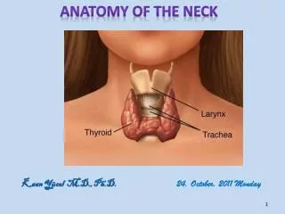
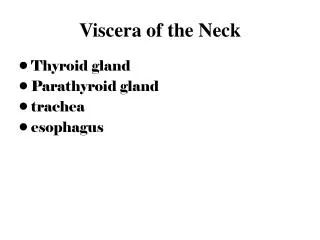
Compartments of the neck Neck has 4 major compartments • Vertebral compartment • Cervical vertebrae and associated muscles • Visceral compartment • Parts of respiratory and digestive tracts, important glands (thyroid, parathyroid and thymus) • Two vascular compartments • Contains major blood vessels and vagus nerve
Cervical fascia Superficial Contains platysma muscle, cutaneous nerves, superficial veins and superficial LNs Deep
Deep cervical fascia • Consists of 4 parts • Investing layer • Encircles the neck completely • Pretrachial fascia • Encircles the visceral compartment • Prevertebralfascia • Encircles the vertebral compartment • Carotid sheaths • Enclose the two vascular compartments
Investing layer • Splits to enclose the parotid gl. • Superficial layer forms parotid fascia & attached to zygomatic arch Mandible Mastoid p. E.O. protuberance & s. nuchal line Acromian & spine L. nuchae Clavicle Manubrium
Investing layer • Deep layer forms stylomandibular ligament between styloid process & angle of mandible Stylomandibular ligament Mandible Mastoid p. E.O. protuberance & s. nuchal line Acromian & spine L. nuchae Clavicle Manubrium
Prevertebral fascia • Lies in front of prevertebral muscles • Extends from skull base to T4 body getting attached to anterior longitudinal ligament • Extends around the subclavian artery and brachial plexus and becomes axillary sheath • Phrenic nerve lies behind • Sympathetic trunk lies in front • Cutaneus branches of Cx plexus pierce it
Vagus nerve Anterior Carotid sheath
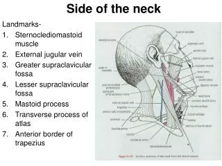
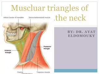
Posterior triangle - contents • External jugular vein after piercing the investing layer. • Accessory nerve • Branches of Cx plexus • Lesser occipital • Great auricular • Transverse cervical • Supraclavicular
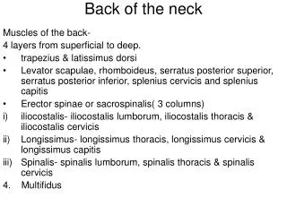
Floor of the posterior triangle • Prevertebral fascia lying on the muscles • Semispinalis capitis • Splenius capitis • Levator scapulae • Scalenus medius • Scalenus anterior
Anterior triangle of the neck • Suprahyoid muscles • Digastric: poterior belly by facial N. & anterior belly by nerve to mylohyoid • Stylohyoid: by facial nerve • Mylohyoid: by nerve to mylohyoid, a branch of inferior alveolar nerve (mandibular division) • Geniohyoid: by C1 through hypoglossal nerve
Anterior triangle…. • Infrahyoid muscles • Sternohyoid • Omohyoid • Thyrohyoid • Sternothyroid These are depressors of the larynx. Supplied by ansa cervicalis (C1-C3)
Carotid triangle • Tip of the greater horn of hyoid is an important reference point • Note the relationships of hypoglossal nerve
Root of the neck • All structures that pass between the head and thorax or upper limb and thorax must pass through the root of the neck
Subclavian Artery • 1st part of subclavian artery • Vetebral A • Internal thoracic A • Thyrocervical trunk • 2nd part of subclavian artery • Costocervical trunk • 3rd part of subclavian artery • Dorsal scapular A
Thoracic duct in the neck • Join the junction between the left subclavian and internal jugular veins