Download
1 / 39
390 likes | 742 Views
CaseA 56 year old man comes to your office complaining of three months of progressive fatigue and dyspnea on exertion. Several times in the past month he has awakened from sleep with severe breathlessness and felt a need to sit up in order to breath. He denies any chest pain or pressure. He also has noticed some ankle swelling. He has no past medical history of heart disease, hypertension or diabetes. His family history is negative for heart disease. He does not smoke and drinks alcohol only r22

E N D
1. Heart Muscle Disease:Cardiomyopathy Laura Wexler, M.D.
558-5575
wexlerl@ucmail.uc.edu
2. Case
A 56 year old man comes to your office complaining of three months of progressive fatigue and dyspnea on exertion. Several times in the past month he has awakened from sleep with severe breathlessness and felt a need to sit up in order to breath. He denies any chest pain or pressure. He also has noticed some ankle swelling. He has no past medical history of heart disease, hypertension or diabetes. His family history is negative for heart disease. He does not smoke and drinks alcohol only rarely. He takes no medications.
3. Physical Exam BP 105/70, P 98 regular, T 98.6?, RR 20
Carotids are low volume with normal upstroke.
JVP elevated: 10 cm above the sternal angle.
Lungs: Bibasilar rales.
Heart: PMI diffuse, palpable at the anterior axillary line.
S1 diminished intensity, S2 normal, S3 is present.
2/6 holosystolic murmur at the apex.
Abdomen: Liver is enlarged (span 11 cm) and slightly tender to pressure. Positive hepatojugular reflex (+HJR). No ascites.
Extremities: Mild edema of both feet and ankles.
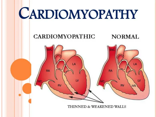
4. Dilated Cardiomyopathy Dilation of one or both ventricles
Globally impaired ventricular systolic function: both ventricles or predominantly the left ventricle. Isolated RV cardiomyopathy is rare.
5. Cardiomyopathies
6. Diagnostic studies ECG: NSR at 82 bpm. No specific findings
Imaging
Chest X-Ray: cardiomegaly and pulmonary congestion.
Echocardiogram: Biventricular enlargement and global hypokinesis.
Radionuclide ventriculogram (MUGA): RVEF 30%, LVEF 20%, global hypokinesis.
Cardiac cath: contrast left ventriculogram. *
7. Dilated Cardiomopathy: MUGA
8. Systolic heart failure
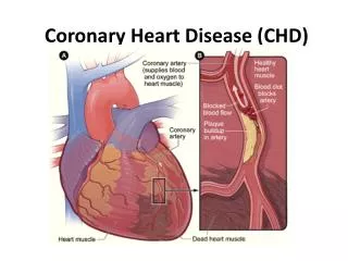
9. Etiology of dilated cardiomyopathy Coronary artery disease
Idiopathic
Hypertensive heart disease
Familial/genetic
Viral/other infectious agents (HIV)
Immune/autoimmune
Alcoholic/toxic (cocaine, chemotherapeutic drugs)
Infiltrative (hemochromatosis, sarcoidosis, amyloidosis)
Post partum
10. Natural History of Dilated Cardiomyopathy Congestive heart failure
Arrhythmias (Afib, VT)
Sudden death
Thromboembolism
Chest pain
11. Diagnosis of Dilated Cardiomyopathy
Exclude other causes of contractile failure
(HTN, CAD, valvular disease).
Test for specific etiologies
?Percutaneous endomyocardial biopsy
12. Goals of Therapy in Dilated Cardiomyopathy Alleviate symptoms of dyspnea
Improve exercise tolerance
Prevent progressive cardiac dilation (remodeling)
Prolong survival
13. Case
A 19 year old college freshman collapses on the basketball court during practice. Despite prompt bystander initiated CPR and the arrival of paramedics within 4 minutes, multiple attempts at defibrillation and prolonged ACLS are unsuccessful and he is pronounced dead at a nearby hospital. He has no history of ill health, syncope or dizzy spells and never used illicit drugs. What is his autopsy likely to show? *
14. Cardiomyopathies
15. Hypertrophic Cardiomyopathy Left ventricular hypertrophy
Myofibrillar disarray
Normal or supernormal contractile function
Impaired diastolic function: impaired diastolic relaxation and decreased LV compliance
16. Cardiac physiology
17. Natural History of Hypertrophic Cardiomyopathy Dyspnea on exertion
Chest pain
Syncope
Sudden death
18. Etiology of Hypertrophic Cardiomyopathy Mutations in sarcomeric contractile protein genes
?-myosin heavy chain, cardiac troponin T and I, ?-tropomyosin, cardiac myosin binding protein C, essential light chain, myosin regulatory light chain
Familial (autosomal dominant with variable penetrance) or sporadic
Some mutations are associated with particularly high risk of sudden death
19. Diagnosis: Physical Findings in Hypertrophic Cardiomyopathy JVP: Prominent �a� wave
PMI: LV heave, double apical impulse (palpable �a� wave)
Heart sounds: Loud S4
20. Diagnostic Tests in Hypertrophic Cardiomyopathy ECG: LVH with �strain� pattern
Chest Xray: Usually normal
Imaging:
Echocardiogram
Radionuclide ventriculogram
Contrast left ventriculogram
21. ECG: LVH with �strain� pattern
22. Hypertrophic Cardiomyopathy
23. Hypertrophic cardiomyopathy *
24. Hypertrophic obstructive cardiomyopathy
25. Hypertrophic Obstructive Cardiomyopathy (HOCM)aka Idiopathic Hypertrophic Subaortic Stenosis - (IHSS) Asymmetric septal hypertrophy
Dynamic systolic obstruction of left ventricular outflow: apposition of the bulging septum and the anterior leaflet of the mitral valve
26. Hypertrophic obstructive cardiomyopathy
27. Physical Exam in HOCM Brisk early carotid impulse
�Triple ripple� PMI: palpable �a� wave, followed by double systolic impulse
�Dynamic� systolic ejection murmur: changes with changes in LV volume or contractility.
28. Dynamic murmur of HOCM Smaller LV volume brings septum closer to anterior MV leaflet: more obstruction and louder murmur.
Larger LV volume separates upper septum from anterior MV leaflet: less obstruction and softer murmur.
29. How to alter LV volume Increase LV volume
Squatting
Passive leg lifting
Slow heart rate
IV volume infusion
Decrease LV volume
Stand (after squatting)
Valsalva maneuver
Increase heart rate
Amyl nitrate
Volume depletion
30. Hypertrophic Cardiomyopathy:Management Predict risk of sudden death:
Early age at presentation
Positive family history
Massive hypertrophy: LV >35 mm
Syncope
Non-sustained VT on Holter
Genetic typing
Prevent sudden death
Internal cardiac defibrillator (ICD)
31. Hypertrophic Cardiomyopathy: management Enhance impaired LV diastolic function (improve filling)
Slow heart rate
Maintain normal sinus rhythm
[Drugs to enhance myocardial relaxation]
Reduce obstruction caused by septal/mitral valve apposition:
Avoid dehydration and vasodilators
Negative inotropic drugs (beta blockers, disopyramide)
Surgical septal myectomy
Dual chamber (atrial and ventricular) pacemaker
32. Hypertrophic cardiomyopathy: pathophysiology
33. Restrictive Cardiomyopathy Abnormally stiff myocardium:
Fibrosis, infiltration, idiopathic
Impaired diastolic function
(Usually) preserved systolic function
34. Restrictive Cardiomyopathy Pathophysiology
Impaired biventricular filling
Elevated right and left atrial pressures
Symptoms: Dyspnea, exercise intolerance
Signs
Increased JVP (large �a� wave), edema, ascites
Increase JVP with inspiration
(Kussmaul's sign)
35. Restrictive Cardiomyopathy Diagnosis:
Cardiac catheterization:
Restricted filling pattern during diastole
RV biopsy
36. Restrictive physiology
38. Dynamic outflow gradient: IHSS
39. Restrictive Cardiomyopathy: Pathophysiology