Download

1 / 64
730 likes | 1.1k Views
ELECTROCHEMICAL DNA BIOSENORS. Prof. Mehmet OZSOZ Ege University, Faculty of Pharmacy, Dept. of Analytical Chemistry, 35100 Bornova / IZMIR ozsozs@pharm.ege.edu.tr. SUMMARY. What’s a biosensor? Electrochemical DNA Hybridization Sensing Strategies

E N D
ELECTROCHEMICAL DNA BIOSENORS Prof. Mehmet OZSOZ Ege University, Faculty of Pharmacy, Dept. of Analytical Chemistry, 35100 Bornova / IZMIR ozsozs@pharm.ege.edu.tr
SUMMARY • What’s a biosensor? • Electrochemical DNA Hybridization Sensing Strategies • Inosine based hybridization detection by using carbon paste electrode (CPE) • Gold nanoparticles based detection of hybridization by using disposable pencil graphite electrode (PGE) • Detection of Factor V Leiden Mutation by using CPE and PGE from real PCR samples. • Carbon Nanotubes • TiO2 nanoparticles
Introduction • The detection of specific DNA sequences provides the basis for detecting a wide variety of infectious and inherited diseases. • Traditional methods for DNA sequencing, based on the coupling of electrophoretic separations and radioisotopic detection, are labor intensive and time consuming, and are thus not well suited for routine and rapid medical analysis, particularly for point-of-care tasks.
Electrochemical hybridization biosensors (genosensors) for the detection of DNA sequences may greatly reduce the assay time and simplify its protocol. Such fast on-site monitoring schemes are required for quick preventive action and early diagnosis. • Therefore, genosensors have recently been the subject of extensive research activities.
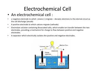
Basic principle of a glucose biosensor GOX -D-glucose + O2 + H2O Gluconolactone + H2O2 Transducer Analytical signal
Electrochemical DNA Hybridization Sensing Strategies 1.Label based a) Hybridization indicators • metal complexes • organic dye molecules • anticancer agents etc. b) Labelled probe • Metal label (Au or Ag-nanoparticles,) • oligonucleotide containing -SH, -NH2, groups. 2. Label free • Electrochemical signals of DNA purine bases guanine, (Inosine), adenine
Inosine is an electro-inactive analogue of guanine, which can also bind to cytosine by forming two hydrogen bonds.
Oxidation signal of DNA bases Guanine, Adenine Inosine,Adenine
Electrochemical DNA biosensors were described for the electrochemical DNA detection procedure based on oxidation signals of guanine and Au nanoparticles to detect an inherited disease; Factor V Leiden Mutation using polymerase chain reaction (PCR) amplicons and synthetic oligonucleotides.
The Factor V Leiden mutation, • designated as 1691 G > A or R506Q, is the major heritable risk factor for venous thromboembolism. • This mutation in the coagulation factor V gene results in the resistance of Factor V to inactivation by activated protein C (APC). • If the coagulation Factor V cannot be inactivated, blood coagulates in venums.
Sequences Wild-type (WT) capture probe : 5’ – AAT ACC TIT ATT CCT CIC CTI TC – 3’ Wild-type target : 5’ – GAC AGG CGA GGA ATA CAG GTA TT – 3’ Mutant (MT) capture probe : 5’ – AAT ACC TIT ATT CCT TIC CTI TC – 3’ Mutant target : 5’ – GAC AGG CAA GGA ATA CAG GTA TT – 3’
Part I • An electrochemical DNA biosensor was described for the detection of Factor V Leiden mutation and the discrimination of mutation type using the oxidation signal of guanine in connection with DPV for the first time. • There have not yet been any literature reports about the detection of heterozygous or homozygous mutations from PCR amplified amplicons by using the guanine signal without any modifications in the native bases or any external labels.
In this study, • Inosine substituted synthetic oligonucleotide capture probes related to the wild – type or mutant type amplicons were used and these probes were hybridized with their complementary DNA sequences (target sequence or PCR amplicons) at carbon paste electrode (CPE).
YES / NO SYSTEM for hybridization detectionNo signal is observed from inosine modified probe.After hybridization, a signal is derived from the guanine bases in the target.
Experimental • CPE Activation :1.7V 60 sec. in 0.05M phosphate buffer solution (PBS). • Inosine-labelled probe immobilization :+0.5V 300s. in acetate buffer solution(ABS). • Washing step with ABS. • Hybridization with the synthetic target or PCR sample : Capt. probe modified CPE was inverted and ~10µl of the target/ denatured PCR amplicon (heating in a water bath at 950C for 6 min. and subsequent freezing in ice bath for 2 min.) was pipetted directly onto the capture probe. • Washing step %1 SDS buffer 3s and then immediately dipped into blank Tris-HCl buffer solution(TBS). • Measurement : The oxidation signal of guanine was measured by using differential pulse voltammetry (DPV) in blank ABS by scanning from-0.80-+1.40V.
Experimental Procedure • When hybridization was occured between probe and target on CPE surface, a guanine oxidation signal at ~+1.00 V was appeared. The YES / NO system was established for the electrochemical detection of allele – specific mutation on Factor V.
Carbon Nanotubes(CNT) • Multi walled carbon nanotubes (MWNTs) were used as nanowires which combined DNA molecules to a carbon paste electrode(CPE) • Unique electronic and mechanical properties and chemical stability • CNT accelerate the electron transfer
DNA-Directed Attachment of Carbon Nanotubes for EnhancedLabel-Free Electrochemical Detection of DNA Hybridization
Part II Electrochemical Genosensor based on colloidal gold nanoparticles
Gold nanoparticleshave been an attractive material in research for a long time … Mirkin, C. A.; Letsinger, R. L.; Mucic, R. C.; Storhoff, J. J. Nature1996, 382, 607.
The visible color shift and aggregation of oligonucleotide modified Au nanoparticles upon binding to target DNA is a well-described event. Color shift is only observed from the hybridization with the target DNA. Elghanian, R.; Storhoff, J. J.; Mucic, R. C.; Letsinger, R. L.; Mirkin, C. A. "Selective Colorimetric Detection of Polynucleotides Based on the Distance-Dependent Optical Properties of Gold Nanoparticles," Science, 1997, 277, 1078-1080.
Nanoelectrodes with nanoparticles Hybridization forms a self-assembly of Au nanoparticles in the nanogap between two nanoelectrodes. Silver precipitation on Au nanoparticles facilitates the electrical flow from one electrode to the other. Park, S.-J.; Taton, T. A.; Mirkin, C. A. "Array-Based Electrical Detection of DNA Using Nanoparticle Probes," Science, 2002, 295, 1503-1506.
Our strategy depended on pure electrochemistry of Au nanoparticles : • When hybridization occured between complementary probes conjugated to Au nanoparticles and target on pencil graphite electrode (PGE) surface, Au oxide wave at about 1.20 Vappeared. • The changes in this electrochemical signal was used to detect hybridization.
Specific probes were immobilized onto the Aunanoparticles in two different modes; • a) Inosine substituted probes were covalently attached from their amino groups at 5` end using N-(3-dimethylamino)propyl)-N’-ethylcarbodiimide hydrochloride (EDC) and N-hydroxysulfosuccinimide (NHS) as a coupling agent onto a carboxylate terminated L-cysteine self assembled monolayer (SAM) preformed on the Au nanoparticles and • b) Probes with a hexanethiol group at their 5’ phosphate end formed a SAM on Au nanoparticles.
The base sequences used Synthetic PCR product: 5’ – CCT GCC CCA ATC CCT TTA TTA CCC CCT CCT TCA GAC ACC TCT AAC CTC TTC TGG CTC AAA AAG AGA ATT GGG GGC TTA GGG TCG GAA CCC AAG CTT AGA ACT TTA AGC AAC AAG ACC ACC ACT TCG AAA CC –3’ Thiol-capped probe: 5’ – SH – C6H5 - GGT TTC GAA GTG GTG GTC TTG – 3’ Wild-type (WT) capture probe: 5’ – NH2 - AAT ACC TIT ATT CCT CIC CTI TC – 3’ Wild-type target: 5’ – GAC AGG CGA GGA ATA CAG GTA TT – 3’ Mutant (MT) capture probe: 5’ – NH2 - AAT ACC TIT ATT CCT TIC CTI TC – 3’ Mutant target: 5’ – GAC AGG CAA GGA ATA CAG GTA TT – 3’
Results • For the detection of hybridization between the Factor V Leiden WT or MT capture probe immobilized Au nanoparticles and target DNA, an aliquot of the probe modified Au nanoparticles is simply introduced onto the target immobilized electrode. • The appearance of the Au oxidation signal confirmed the presence of the sought-after DNA sequence.
WT probe with WT target at PGE %R. S. D. = 7.64 % (n=5). MT probe with the MT target at PGE % R. S. D. = 7.42 % (n=5). The detection limits, (S/N=3) 0.78 fmole/mL target with WT probe modified gold nanoparticles 0.83 fmole/mL target with MT probe modified gold nanoparticles.
Histomogram showed that, bare and TiO2 modofied carbon paste electrode(CPE) behaviours, when the probe or hybrid immobilized onto the electrode surface.In the first column, synthetic probe seguence modified (ssDNA) bare CPE, in the second column, synthetic probe seguence modified (ssDNA) TiO2 contained CPE, In the third column, synthetic hybrid modified (dsDNA) bare CPE, In the forth column, synthetic hybrid modified (dsDNA) TiO2 contained CPE. Also the similar results obtained with pencil graphite electrodes.
Future work • For this study, hybridization detection (after finding TiO2 nanoparticles’ attractivity on ss or ds DNA) by using CPE and PGE.
Electrochemical Coding of Single-NucleotidePolymorphisms By Monobase-Modified GoldNanoparticles
Part III Electrochemical Genosensor for the Discrimination of HSV (Herpes Simplex Virus) Type I and II
Herpes Simplex Virus; Type I PCR Product 5’TCAACTTCGACTGGCCCTTCTTGCTGGCCAAGCTGACGGACATTTACAAGGTCCCCCTGGAGACGGGTACGGCCGCATGAACGGCCGGGGCGTGTTTCGCGTGTGGGACATAGGCCAGAGCCACTTCCAGAAGCGCAGCAAGATAAAGGTGAACGGCATGGTGAGCATCGACATGTACGG 3’ Type II PCR Product 5’TCAACTTCGACTGGCCCTTCGTCCTGACCAAGCTGACGGAGATCTACAAGGTCCCGCTCGAGACGGGTACGGGCGCATGAACGGCCGGGGTGTGTTCCGCGTGTGGGACATAGGCCAGAGCCACTTCCAGAAGCGCAGCAAGATAAAGGTGAACGGCATGGTGAACATCGACATGTACGG 3’