Download
1 / 72
730 likes | 1.35k Views
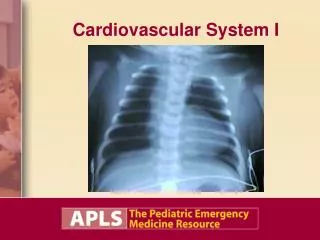
Cardiovascular System I. Objectives. Present the clinical features and emergency management of cardiovascular disorders, including: Recognize congenital and acquired heart disease. Outline management of ductal dependent lesions. Identify patients with myocarditis.

E N D
Objectives • Present the clinical features and emergency management of cardiovascular disorders, including: • Recognize congenital and acquired heart disease. • Outline management of ductal dependent lesions. • Identify patients with myocarditis.
Congenital Heart Disease: Recognition and Stabilization • Rapid cardiopulmonary assessment to recognize and manage life-threatening illness caused by heart disease • Understand the physiology of different conditions to optimize treatment plans.
Critical Concepts • Dysrhythmias can cause serious cardiovascular compromise. • Structural congenital heart disease can present in many different ways at many different ages. • Acquired heart disease can be subtle yet life-threatening.
Case Study 1:“Rapid Breathing” • 10-day-old infant is brought to ED by mother for rapid breathing and not eating well. • Product of normal spontaneous vaginal delivery • Spent 2 days with mother in hospital • Uneventful course, including circumcision • Birth weight 3.2 kg
Case Study 1 (continued) • Slow to breastfeed since birth • Would gasp and cry after sucking for a short time. Difficulty feeding. • 3 to 4 wet diapers per day • No congestion, no fever • No vomiting with feedings • 2 yellow seedy stools since passing meconium after birth
Initial Assessment (1 of 2) PAT: • Abnormal appearance, abnormal breathing, abnormal circulation Vital signs: • HR 170, RR 70, BP 82/40, T 37°C (rectal), Wt 3.4 kg, O2 sat 90% on room air
Initial Assessment (2 of 2) A: No evidence of obstruction B: Elevated RR and labored C: Pale, diaphoretic, tachycardia, weak pulse, cyanosis D: GCS grossly normal but in distress and inconsolable E: No signs of head injury, fractures, or bruising
Detailed Physical Exam • Lung sounds equal bilaterally with rales in both bases • Hyperactive precordium with a gallop rhythm • Pulses weak in distal and lower extremities • Distended abdomen with liver palpable 4 cm below right costal margin
Question What is your general impression of this patient?
General Impression • Impending cardiopulmonary failure (compensated shock) • Cyanosis, diaphoresis • Pale, tachycardia What are your initial management priorities?
Management Priorities (1 of 3) • ABCs • Give 15L O2 by nonrebreather mask or 100% O2 by BMV, or perform endotracheal intubation. • Start IV, obtain blood glucose. • ECG and monitor rhythm on cardiac monitor • CXR • Administer fluid challenge: 10 cc/kg NS
Management Priorities (2 of 3) • Administer prostaglandin E1 (PGE1): • 0.05 to 0.1 mcg/kg/min • Intubate to protect against apnea and relieve stress from work of breathing. • Consider furosemide (0.5 to 1 mg/kg). • Sepsis work-up and then antibiotics • Defer lumbar puncture.
Management Priorities (3 of 3) • Cardiology consultation or transfer to pediatric cardiology center emergently: • Echocardiogram • If blood pressure and perfusion do not improve, add inotropic agent: • Dobutamine: 2 to 20 mcg/kg/min • Epinephrine: 0.1 to 1.5 mcg/kg/min
Case Discussion (1 of 2) • This infant is in CHF. • Poor feeding and easy fatigability • Gallop rhythm and enlarged liver • Diminished pulses • Shock: • Altered mental status, compensated shock (tachycardia, diaphoresis, respiratory distress, normal BP in upper extremities)
Case Discussion (2 of 2) • Possible ductal dependent lesion: • Right age for presentation of shock triggered by closure of the ductus arteriosus • Measure blood pressure in four extremities • Assess oxygenation response to supplemental oxygen
Case Progression: Version 1 • BP differential noted in lower extremities. • Oxygenation improves to 99% with supplemental oxygen. • CXR shows cardiomegaly and pulmonary edema. • Echocardiogram demonstrates coarctation of the aorta. • Infant improves with PGE1 infusion, diuretics, and inotropes.
Case Progression: Version 2 • Oxygenation fails to improve with supplemental oxygen (remains 90%). • Oxygenation declines further to <80%. • CXR is nonspecific. • Echocardiogram demonstrates transposition of the great vessels. • Infant improves with PGE1 infusion. • Surgical intervention is scheduled.
Background: Structural Congenital Heart Disease • Congenital heart disease: 5 to 8 cases per 1,000 live births • Child with congenital anomaly usually does not show cardiovascular problems in utero. • Changes at birth place great stress on infant’s cardiovascular system. • Some cyanotic heart conditions are highly dependent on shunting through ductus arteriosus. Closure can be terminal event.
Clinical Features: Your First Clue • Age • Progressive deterioration (mild) followed by suddenly progressing to critical condition • Cyanosis • Congestive Heart Failure (CHF) • Consider concurrent sepsis
Diagnostic Studies (1 of 3) • Radiology: • Pulmonary hypoperfusion: pulmonic stenosis, TOF, TA • CHF (if large VSD present to allow high-output failure, e.g., increased right-sided flow) • Some classic CXR appearances (more classic if condition is permitted to worsen): • TGA: Egg on side • TAPVR: Snowman • TOF: Boot shaped
Diagnostic Studies (2 of 3) • ECG: • Right axis (RVH): Normal for newborns • Left axis: Hypoplastic right heart, tricuspid atresia, endocardial cushion defect (AV canal) • ST-T changes, strain, ischemia • Dysrhythmia • Prolonged QT • Low voltage
Diagnostic Studies (3 of 3) • Laboratory: • Glucose: Any child in distress needs to have hypoglycemia excluded. • CBC: Look for anemia, signs of sepsis. • Electrolytes: Congenital adrenal hyperplasia, salt-wasting form • Arterial blood gas: Hyperoxia text
Fetal Circulation (1 of 2) • Placenta oxygenates blood and returns to right atrium (RA) via IVC. • Preferentially shunts across FO to LA. • LV ejects most oxygenated blood to carotids and coronaries.
Fetal Circulation (2 of 2) • Superior vena cava (SVC) returns deoxygenated blood to RA where it mixes with oxygenated blood from the placenta. • Preferentially enters RV. • RV ejects into PA. • No pulmonary capillary flow, so PA is shunted into the descending aorta via the ductus arteriosus.
Differential Diagnosis: What Else? (1 of 2) • Other cyanotic and acyanotic congenital structural heart disease • Ductal dependent coarctation • Hypothermia • Sepsis • TORCH
Differential Diagnosis: What Else? (2 of 2) • Congenital adrenal hyperplasia (CAH) • Hypoglycemia • Shaken baby syndrome/intracranial lesion • Catastrophic gastrointestinal process, e.g., volvulus
Normal CV System Function • Represented by vital signs (O2 sat included) • Factors affecting cardiac output (perfusion): • Heart rate • Stroke volume • Contractility • Vascular resistance • Children <8 years predominantly increase their HR to increase cardiac output (unable to increase stroke volume until >10 years).
Normal Vital Signs For Age HR RR BP (systolic) Newborn 90-180 40-60 60-90 1 month 110-180 30-50 70-104 3 months 110-180 30-45 70-104 6 months 110-180 25-35 72-110 1 year 80-160 20-30 72-110 2 years 80-140 20-28 74-110 4 years 80-120 20-26 78-112 6 years 75-115 18-24 82-115 8 years 70-110 18-22 86-118 10 years 70-110 16-20 90-121 12 years 60-110 16-20 90-126 14 years 60-110 16-20 92-130
Transition from Fetal Circulation • Placental circulation is interrupted at birth: • Increase in systemic arterial blood pressure • Spontaneous respirations • Decreased pulmonary vascular resistance, increasing pulmonary blood flow • Foramen ovale closes. • Ductus arteriosus closes. • This initial rapid change slows down over first 24 hours of life.
Cyanotic Heart Disease (CHD) • Cyanotic: Refractory to oxygen • Right to left shunting • Some lesions (e.g., TGA) are highly dependent on a shunt (VSD, PDA) • Cyanosis usually presents shortly after birth.
Cyanotic CHD • 5 Ts: • Truncus arteriosus • Tetralogy of Fallot (TOF) • Transposition of the great arteries (TGA) • Tricuspid atresia • Total anomalous pulmonary venous return (TAPVR) • Severe aortic stenosis • Hypoplastic left heart • Severe coarctation of the aorta
Tetralogy of Fallot (TOF) • Pulmonic stenosis • Aortic override • VSD • RVH • Right-to-left shunting through VSD dependent on severity of pulmonic stenosis
Tricuspid Atresia • RV is hypoplastic. • Right-to-left shunt through VSD
Cyanosis • Respiratory disorder • Hemoglobin disorder • Acrocyanosis (normal newborns): Cold stress and peripheral vasoconstriction • Generalized or central cyanosis often due to cyanotic congenital heart disease. Often worsened by crying.
Hyperoxia Test • Administer 100% oxygen. • Significant increase in PaO2 seen with pulmonary and hemoglobin disorders. • In CHD, PaO2 will not increase or it will increase slightly. • Deoxygenated blood bypasses lungs and goes directly to left side of heart, diluting the fully oxygenated blood coming from lungs with deoxygenated blood.
CHD • Increased pulmonary vascularity: • Total anomalous pulmonary venous return • Truncus arteriosus • Transposition of the great arteries • Other complex lesions without pulmonic stenosis • Decreased pulmonary vascularity: • Tetralogy of Fallot • Ebstein’s anomaly • Hypoplastic right heart, tricuspid atresia • Complex lesions with pulmonic stenosis
Prostaglandin E1 • Keeps the ductus open • 0.05 to 0.1 mcg/kg/min with an increase to 0.2 mcg/kg/min over several minutes • Side effects: Apnea, pulmonary congestion, fever, hypotension, seizures, and diarrhea • Consider elective intubation.
Noncyanotic CHD • May present with CHF or heart murmurs heard during physical exam • Left-to-right shunts • Excess pulmonary vascularity • ASD, VSD, PDA • Obstructive lesions • Aortic stenosis, coarctation of the aorta, mitral stenosis, pulmonic stenosis
Clinical Features • CHF: Tachypnea, tachycardia, diaphoresis, decreased feeding, hepatomegaly, murmurs, gallop rhythms, pulmonary edema • Decreased activity or poor sleeping with respiratory distress
Diagnostic Studies • CXR: Cardiomegaly, pulmonary vascular congestion • ECG: Abnormal axis, ST segment changes • Echocardiogram: Definitive anatomic diagnosis, degree of congestive heart failure (chamber sizes, contractility)
Management of CHF • Give oxygen, assisted ventilation if needed. • Elevate head and shoulders 45 degrees. • Monitors, pulse oximetry • Obtain IV access. • Send laboratories. • CXR and ECG • Furosemide, nitroglycerin, digoxin • Inotropes (dobutamine) for signs of shock
Case Study 2:“Chest Pain, SOB” • 10-year-old boy presents with chief complaint of chest pain and shortness of breath. • 5 days of cold and cough symptoms • He has been lying around a lot and has missed 1 week of school. • Usually a very active child but complains that he is “just too tired” to play
Initial Assessment PAT: • Abnormal appearance, abnormal breathing, abnormal circulation Vital signs: • HR 130, RR 44, BP 90/65, T 37.8°C, O2 sat 90% on room air, increases to 100% on O2
Initial Assessment A: Patent B: Intermittently shallow and deep; rapid respiratory rate C: Pale; pulse rapid, thready, and weak D: No focal deficits, GCS 15 E: No signs of injury