Download
1 / 54
750 likes | 1.7k Views
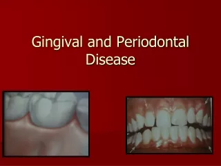
EPIDEMIOLOGY OF PERIODONTAL DISEASE. ASPECT OF NORMAL GINGIVA. CLINICAL NORMAL GINGIVA. HEAVILY PIGMENTED GINGIVA. Supragingival calculus is depicted on the buccal surfaces of maxillary molars adjacent to orifice for Stenson’s duct.

E N D
Supragingival calculus is depicted on the buccal surfaces of maxillary molars adjacent to orifice for Stenson’s duct
Extensive supragingival calculus on the lingual surfaces of lower anterior teeth
Extensive supragingival calculus is on the lingual surfaces of lower anterior teeth
Dark pigmented deposits of subgingival calculus on the distal root of an extracted lower molar.
A lower incisor depicting a prominent root without any attached gingiva and accompanying gingival recession
A patient following the placement of a soft tissue graft to gain attached gingiva and treat the gingival recession
Overhanging margin of restoration and atrophied and inflamed gingival papilla
48-hour plaque growth. Generalized gingivitis at the margins of almost all teeth
Heavy calculus deposits on facial surfaces of upper first molar and second premolar.
Clinically normal gingiva- profuse bleeding after 30 seconds.
Extent of pocket revealed by periodontal probe on mesial of central incisor
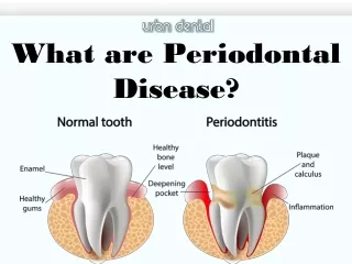
The epidemiology is the science of epidemics. Epidemiological methods are utilized for determining what proportion of the population at a given time is affected by a disease
The purpose of epidemiology is to increase understanding of the disease process to identify the risk factors or deteminants of disease. One of the most valuable employed in dental epidemiology is the epidemiologic index which help in defining diagnosis
A GOOD EPIDEMIOLOGY INDEX : Must be easy to use Permit the examination of many people in a short period of time
TYPES OF DENTAL INDICES 1/ Measures the number or proportion of people in a population with or without a specific condition at a specific point in time or interval of time 2/ Merasures the number of people affected and severity of the specific condition at a specific time or interval of time
INDICES I. Plaque indices II. Gingival indices III. Periodontal indices IV. Treatment needs indices
Plaque indices • Plaque index • Interdental hygiene index • Hygiene index
Plaque index This index concerns thickness of plaque along the gingival margin; this plaque plays role in the etiology of gingivitis Grade Description 0 No plaque 1 Thin film of plaque at the gingival margin, visible Only when scraped with explorer 2 Moderate amount of plaque along the gingival margin; Interdental space free of plaque; Plaque visible with the naked eye. 3 Heavy plaque accumulation at the gingival margin; Interdental space filled with plaque
Gingival Index Grade Description 0 Normal gingival, no inflammation, no discoloration, no bleeding 1 Mild inflammation, slight color change, mild alteration of gingival surface No bleeding 2 Moderate inflammation, erythema, swelling, bleeding on probing, or when Pressure applied 3 Severe inflammation, severe erythema and swelling, tendency toward spontaneous Hemorrhage, some ulceration
Papilla bleeding index Grade Description 1-point 20-30 seconds after probing the mesial and distal sulcus with a periodontal probe, a single bleeding point is observed 2 -line/Points Thin film of plaque at the gingival margin, visible only when scraped with an explorer 3 -Triangle The interdental triangle becomes more or less filled with blood 4 - Drops Profuse bleeding. Immediately after probing, blood flows into the interdental area to cover portions of the tooth or gingiva
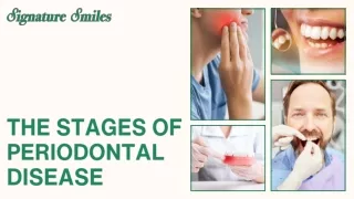
Periodontal Disease Index (PDI) 0 No inflammation, no alterations in the gingiva Gingiva 1 Mild to moderate gingivitis at some locations on the gingival margin 2 Mild to moderate gingivitis of the entire gingival margin 3 Advanced gingivitis with severe erythema, hemorrhage, ulceration Periodontium 4 Up to 3mm of attachment loss, measured from the cementoenamel junction 5 3-6 mm of attachment loss 6 More than 6 mm of attachment loss
COMMUNITY PERIODONTAL INDEX OF TREATMENT NEEDS (CPITN) • Code CPI TN • 0 Healthy • 1 Bleeding on probing I . Oral hygiene instruction • 2 Supra and\or Subgingival • calculus II . I+ calculus removal • Iatrogenic marginal • irritation • 3 Shallow pockets up to • 5 mm III . I+II+ complex treatment • 4 Deeper pockets from • 6mm
The Periodontal Screening and Recording ( PSR ) The PSR examination was developed in order to streamline the data gathering and record keeping for the screening periodontal examination
The PSR exam is patterned after the Community Periodontal Index of treatment needs (CPITN ) of the world health organization
This exam is completed with periodontal probe that has a ball at the tip and a black or colored band from 3.5-5.5mmWhile six sites are examined per tooth
PSR Codes • Code 0 : The deepest probing in the sextant is < 3.5mm The colored band on the probe remains completely visible There is no bleeding, calculus, or defective restorations The patient needs preventive care only
Code 1:The colored band on the probe remains visibleThere is no calculus or defective restorative margins There is bleeding at the gingival marginPlaque must be removed and the patient instructed in proper oral hygiene
Code 2: The colored band on the probe remains completely visibleThere is detectable calculus and or defective restorative marginsTreatment consists of plaque and calculus removal, Correction of plaque retentive factors and oral hygiene instruction