Download
1 / 1
10 likes | 82 Views
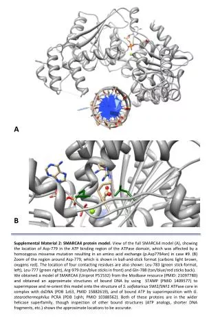
Detailed model view of SMARCA4 protein showing impact of missense mutation at Asp-779 based on structural analysis with other proteins. The study provides insights into the binding region and adjacent residues. Model obtained from Modbase with DNA structures aligned using STAMP. Comparison with ATPase core and bound ATP structures reveals evolutionary relationships.

E N D
A B Supplemental Material 2: SMARCA4 protein model. View of the full SMARCA4 model (A), showing the location of Asp-779 in the ATP binding region of the ATPase domain, which was affected by a homozygous missense mutation resulting in an amino acid exchange (p.Asp779Asn) in case #9. (B) Zoom of the region around Asp-779, which is shown in ball-and-stick format (carbons light brown, oxygens red). The location of four contacting residues are also shown: Leu-783 (green stick-format, left), Leu-777 (green right), Arg-979 (tan/blue sticks in front) and Gln-788 (tan/blue/red sticks back). We obtained a model of SMARCA4 (Uniprot P51532) from the Modbase resource (PMID: 21097780) and obtained an approximate structures of bound DNA by using STAMP (PMID 1409577) to superimpose and re-orient this model onto the structure of S. solfataricusSWI2/SNF2 ATPase core in complex with dsDNA (PDB 1z63, PMID 15882619), and of bound ATP by superimposition with G. stearothermophilusPCRA (PDB 1qhh; PMID 10388562). Both of these proteins are in the wider helicase superfamily, though inspection of other bound structures (ATP analogs, shorter DNA fragments, etc.) shows the approximate locations to be accurate.