Download

1 / 66
660 likes | 862 Views
Introduction to Biomolecular Structure and Modeling. Dhananjay Bhattacharyya Biophysics Division Saha Institute of Nuclear Physics Kolkata dhananjay.bhattacharyya@saha.ac.in. Biomolecular Structures. These are determined experimentally by X-Ray Crystallography

E N D
Introduction to Biomolecular Structure and Modeling Dhananjay Bhattacharyya Biophysics Division Saha Institute of Nuclear Physics Kolkata dhananjay.bhattacharyya@saha.ac.in
Biomolecular Structures These are determined experimentally by • X-Ray Crystallography • Nuclear Magnetic Resonance Spectroscopy • Neutron Diffraction Study • Raman Spectroscopy And also by theoretical methods
Nucleic Acid Backbone is Connected to Either of Four Different Bases
C A G T
A-DNA Z-DNA B-DNA
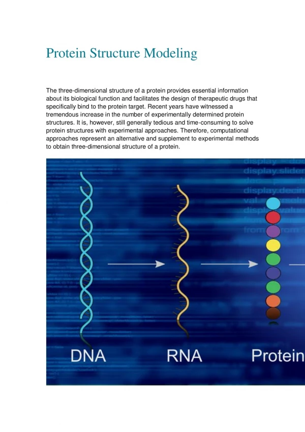
Proteins (polymers) are made up of Amino Acids (monomer units) • There are Twenty different Amino Acids • with different shape, size and electrostatic • properties. • These amino acids form covalent • bonds to form a linear polypeptide chain.
Alanine Phenylalanine Serine Cystine
Glutamic Acid (Negatively charged) Arginine (Positively charged)
Amino Acids are joined together by covalent bonds, called peptide bond, which is structurally very important
a-helix: Hydrogen bonding between every i i+4 residues
b-sheet: Hydrogen bonding between ij, i+1j-1 (Antiparallel), or ij, i+1j+1 (parallel)
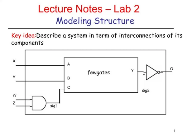
Coordinate System: • External coordinates, such as (x,y,z), (r,q,f), (r,f,z) • Internal coordinates (BondLength, BondAngle, TorsionAngle)
Torsion Angle Bond Angle Bond Length
Generated coordinates • H 0.000000 0.000000 0.000000 • C 0.000000 0.000000 1.089000 • C 1.367073 0.000000 1.572333 • C 2.050610 -1.183920 1.089000 • C 3.417683 -1.183920 1.572333 • H -0.513360 0.889165 1.452000 • H -0.513360 -0.889165 1.452000
y f
Theoretical Modeling of Biomolecules: • Quantum Mechanics based Methods • Statistics based Methods • Classical or Molecular Mechanics methods
Peptide modeling initiated in India by G.N. Ramachandran (1950s) • Postulates: • Impenetrable spherical volumes for each atom • Radius of the sphere depend on atom type • No two atomic spheres can overlap if they are not covalently bonded y f
Between H N O C P S H 2.0 (1.9) 2.4 (2.2) 2.4 (2.2) 2.4 (2.2) 2.65(2.5) 2.65(2.5) N 2.7 (2.6) 2.7 (2.6) 2.9 (2.8) 3.2 (3.1) 3.1 (3.0) O 2.7 (2.6) 2.8 (2.7) 3.2 (3.1) 3.1 (2.9) C 3.0 (2.9) 3.4 (3.2) 3.3 (3.1) P 3.5 (3.3) S Normal and Extreme Limit (within parenthesis) distances (Å) used by Ramachandran co-workers
Fully Allowed Regions Partially Allowed Regions Original Ramachandran Plot
Ramachandran plot for 202 proteins at 1.5A or better resolution
Variation of angle by 5o allowed to fit observed phi-psi of protein structures.
Schrodinger Equation: Quantum Mechanics Time dependent (3 Dimensional) Time independent
DFT formalism with B3LYP Pseudoeigenvalue equation: where Potential due to exchange-correlation, is defined by with a, b and c as parameters obtained from fit with experimental data for sample compounds, Ex are for electron exchange and Ec are for correlation. Essentials of Computational Chemistry by C.J. Cramer (2002) John Wiley & Sons Ltd,
Input data (atom coordinates, basis sets) Generate input guess density (overlap integrals) Construct the potential and Solve Kohn-Sham equation Repeat the cycle using the output density as the input density Generate output densities from Solutions to Kohn-Sham equations NO YES Are input and output density same? Analyze electronic population FLOW CHART DESCRIBING THE DFTMETHODOLOGY
G:C W:W C DE = -26 kcal/mol A:U W:W C DE = -14 G:U W:W C DE = -15 A:G H:S T DE = -10 Strengths of different H-bonds from 33 non-canonical Base Pairs A:G s:s T DE = -6 A:U H:W T DE = -13 A:A H:H T DE = -10 G:A W:W C DE = -15 G:A S:W T DE = -11 A:A W:W T DE = -12 A:U W:W T DE = -13 A:A H:W T DE = -11 2=>NH..N 2=>NH..O 1=>NH..N 1=>NH..O 1=>NH..N 2=>NH..O 1=>NH..O 1=>NH..N 1=>NH..N 1=>CH..O 1=>NH..O 1=>NH..N 2=>NH..N 2=>NH..N 1=>NH..O 1=>NH..N 1=>NH..O 1=>NH..N 2=>NH..N
Considered Energy components, ENHO, ENHN, etc are additive. Additional stabilities, di may come from van der Waals, dipole-dipole etc interactions. Least Squares Fit indicates di, errors should be smallest for best Fit A. Roy, M. Bhattacharyya, S. Panigrahi, D. Bhattacharyya, (2008) J. Phys. Chem. B (in press)
Netropsin like drugs bind in the B-DNA narrow and deep minor groove
Actinomycin D like drugs make their place in between two stacked base pairs by distorting the DNA double helix
DNA kinks by 90o at the dyad location while binding to two subunits of Catabolite Activator Protein (CAP)
TATA-box binding protein transforms the interfacing DNA region to A-DNA like structure
DNA Smooth Curvature induced by Histone proteins in Chromatin (Nucleosome)
Calculation of Base Pair parameters by NUPARM Local Step Parameters: Mean Local Helix Axis: Zm = XmYm, where Xm = Xaxis1 + Xaxis2 and Ym = Yaxis1 + Yaxis2 M is Base Pair Center to Center Vector Tilt : 2.0 * sin-1 ( -ZmY1) Roll: 2.0 * sin-1 ( ZmX1) Twist: cos-1 (( X1Zm) ( X2Zm)) Shift (Dx) MXm Slide(Dy) MYm Rise(Dz) MZm
Partial list of DNA crystal structures available at http://ndbserver.rutgers.edu bd0001 12: A C C G A C G T C G G T bd0003 12: A C C G G T A C C G G T bd0004 12: C G C G A A T T C G C G bd0006 10: G G C C A A T T G G bd0011 12: C G C A A A T A T G C G bd0014 12: C G C G A A T T C G C G bd0015 10: C C G C C G G C G G bd0017 9: C G CG C G G A G bd0018 11: G C G A A T T C G C G bd0019 12: G G C G A A T T C G C G bd0022 12: A C C G G CG C C A C A bd0023 10: C C A G T A C T G G Bd0024 10: C C G A A T G A G G
Base-Pair Step Size of Database Tilt Roll Twist Rise G:G 37 -0.24 5.80 30.99 3.46 G:C 106 -0.33 -5.37 38.52 3.32 C:G 157 0.66 3.81 36.26 3.46 A:A 116 -0.01 0.67 35.92 3.21 A:T 54 0.20 -0.60 32.76 3.25 T:A 18 -0.02 0.07 40.39 3.30 A:C 20 -0.37 0.97 32.73 3.43 C:A 47 -0.19 2.17 37.75 3.48 A:G 34 0.16 5.34 31.92 3.44 G:A 55 -0.23 0.52 38.40 3.14 Average Structural Parameters from Crystal Structures
Curved DNA models built from Crystal parameters (A3G7)n (A10)n (A6G4)n
Bond Angle Deformation q Deformation from equilibrium value q costs energy. Simplest form of energy penalty is: Eq=1/2 k(q-qo)2
Bonds are also stretchable but at a cost of energy Bond Breaking energy
Ethane (three fold symmetry) Ethiline (two fold symmetry)
Between H N O C P S H 2.0 (1.9) 2.4 (2.2) 2.4 (2.2) 2.4 (2.2) 2.65 (2.5) 2.65 (2.5) N 2.7 (2.6) 2.7 (2.6) 2.9 (2.8) 3.2 (3.1) 3.1 (3.0) O 2.7 (2.6) 2.8 (2.7) 3.2 (3.1) 3.1 (2.9) C 3.0 (2.9) 3.4 (3.2) 3.3 (3.1) P 3.5 (3.3) S Normal and Extreme Limiting (within parenthesis) distances (Å) used by Ramachandran co-workers Minimum Energy position: rijo Interaction between Instantaneous Atomic dipoles and Induced Atomic dipoles
E(Dx, Dy, Dz) E(Dx+1, Dy, Dz) E(Dx+2, Dy, Dz) ….. Search for Conformation with Lowest Energy
Multivariable Optimization: NP-hard Problem • Systematic Grid Search procedure: Impossible, large no. variables • Guided Grid Search: Depends on Choice • Approximate Method based on Taylor series • Newton-Rhapson Method:
Energy Landscape of typical bio-molecules Energy Positional Variables
Conformation 0: Calculate energy (Ei) Alter conformation randomly Calculate energy (Ei+1) Calculate ρ = exp(-(Ei+1-Ei)/kT) If ρ > random no accept the conformation Repeat the procedure Energy Uniformly generated Random numbers are used to accept if exp(-U/kT) > random no and reject otherwise Reject Always Accept Accept
Deterministic Method Molecular Dynamics Verlet Algorithm: