Download
1 / 14
150 likes | 819 Views
Acute Tubular Necrosis (ATN). Dr. Belal Hijji, RN, PhD December 14 & 17, 2011. Learning Outcomes. At the end of this lecture, students will be able to: Explain the common causes of acute tubular necrosis. Identify the four phases of ATN and their characteristics.

E N D
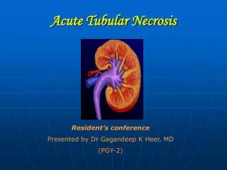
Acute Tubular Necrosis(ATN) Dr. Belal Hijji, RN, PhD December 14 & 17, 2011
Learning Outcomes At the end of this lecture, students will be able to: • Explain the common causes of acute tubular necrosis. • Identify the four phases of ATN and their characteristics. • Discuss assessment and diagnostic findings of ATN. • Identify disease states that increase the risk of ATN. • Discuss the importance of hemodynamic monitoring and fluid balance in a patient with ATN. • Discuss the medical, pharmacological, and nutritional management of ATN. • Discuss the nursing management of a patient with ATN.
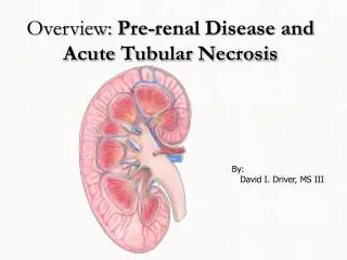
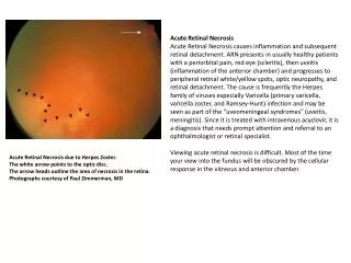
This is the typical appearance of the blood vessels (vasculature) and urine flow pattern in the kidney. The blood vessels are shown in red and the urine flow pattern in yellow.
Causes of ATN • ATN is the most common cause of acute renal failure (ARF); it results from nephrotoxic or ischemic injury that damages the kidney tubular epithelium. • Damage to the kidney cells prevents normal concentration of urine, filtration of wastes, and regulation of acid-base, electrolytes, and water balance. • Ischemic acute tubular necrosis: Ischemic damage occurs as a result of vasodilation associated with sepsis and when hypotension is prolonged (Pre-renal low perfusion). • Toxic acute tubular necrosis: Nephrotoxic damage results from injury by drugs (gentamicin, amikacin, vancomycin) and chemicals (radiopaque dye administered in radiologic diagnostic studies).
Phases of ATN • Initiation phase: This phase is the period from the initial insult until cell injury occurs. It is characterized by acute decrease in GFR (90 - 120 mL/min/1.73 m2) to very low levels, with a sudden increase in serum creatinine and BUN concentrations. This phase lasts hours to days depending on the cause. Prompt treatment can alleviate irreversible damage. • Oliguric or anuric phase: This phase lasts 5-8 days in the non-oliguric patient and 10-16 days in the oliguric (< 500 CC of urine/24 hours) one. The GFR is greatly reduced resulting in high BUN, elevated serum creatinine, hyperkalemia, and metabolic acidosis (rapid breathing, confusion, shock or death in severe cases).
Phases of ATN (Continued….) • Diuretic phase: This phase lasts 7-14 days and is characterised by an increase in GFR and sometimes by polyuria (urine output 2-4 L/day). • Recovery phase: In this phase, kidney tubular cells that have survived prior injury contribute to the healing process through proliferation and migration to the damaged areas. Oliguric or nonoliguric patients may have increased urine output, and kidney function slowly returns to normal or near normal.
Assessment and Diagnosis • Acidosis: pH of <7.35 indicates severe acute kidney insult. • BUN: Increased (normal range 5-25 mg/dL). • Creatinine: Increased (normal range 0.5-1.5). • Creatinine clearance: Decreased (normal range 110-120 mL/min); values <50 mL/min indicates significant kidney dysfunction. • BUN: Creatinine ratio. 20:1 (Ischemia); 10:1 (Toxic).
Disease States That Increase the Risk of ATN • Underlying chronic kidney disease. • Older age, diabetes, and hypertension. • Heart failure. • Respiratory failure. • Sepsis. • Trauma. • Contrast-induced nephrotoxic injury.
Hemodynamic Monitoring and Fluid Balance • Hemodynamic monitoring: This involves surveillance of the central venous pressure (2-5 mm Hg), pulmonary artery occlusion pressure (5-12 mm Hg), cardiac output (SV*HR = 4-8 L/min), and cardiac index (CO/BSA) . Body surface area (BSA) ranges from 2.5 to 4.5 L/min/m2). Stroke volume (SV) is the amount of blood in mL ejected from the ventricles with each beat.
Hemodynamic Monitoring and Fluid Balance (Continued…) • Daily weight and accurate intake and output monitoring. These are powerful indicators of fluid gains or losses over 24 hours. A 1-kg weight gain/ day represents 1 liter of additional fluid retention. • Physical assessment: Signs that suggest extracellular fluid depletion include thirst and decreased skin turgor. Signs that imply intravascular fluid volume overload include pulmonary congestion, increasing heart failure, and high BP.
Medical Management of ATN. Medical treatment of ATN focuses on prevention, fluid balance, medications, and electrolyte balance. • Prevention: This is the only truly effective remedy for acute kidney insult (AKI). Nephrotoxic drugs are avoided in patients with AKI or chronic kidney disease (CKD). Nonsteroidalantiinflammatory (NSAID) drugs are avoided in patients with high creatinine levels. Intravascular contrast dye is delayed, if possible, until the patient is fully rehydrated. • Fluid resuscitation: These include crystalloids (normal saline 0.9%, 0.45% saline, and lactated ringer) and colloids (albumin 5% & 25%). Fluid restriction (1 L/day) aims to prevent circulatory overload and the development of interstitial edema when the kidneys cannot remove excess fluid. Fluid removal is achieved by diuretics. However, hemodialysis is the treatment of choice.
Medical Management of ATN. • Medications: These include diuretics, dopamine (2-3 mcg/kg/min) to increase blood flow to the kidneys. • Nutrition: The recommended energy intake is between 20 and 30 kcal/kg/day with 1.2 to 1.5 g/kg of protein/day to control high BUN. Oral or enteral nutrition is indicated.
Nursing Management of ATN. • Nurses are responsible for recognising potential risk factors for AKI and acts as an advocate for the patient. Such patients include the elderly, dehydrated patients, patients with high creatinine level before admission, and patients for radiologic tests using a dye. • Nurses need to monitor patients for signs of infection (high WBC, redness at wound or intravenous line site, and fever). • Nurses should perform hemodynamic monitoring (HR, BP, CVP, PAOP, CO, and CI), measure weight daily, and record intake and output. Nurses should also recognise early signs and symptoms of fluid overload. • Nurses should monitor the patient for hyperkalemia and hypocalcemia as these increase the risk for cardiac dysrhythmias.
Nursing Management of ATN. • Hyperphosphatemia causes severe itching. Nurses can help by applying emollients for skin care, discouraging scratching, and administering phosphate-binding agents (calcium carbonate & calcium acetate) with food. • Finally, anemia in AKI results from kidney failure to produce the hormone erythropoietin which stimulates bone marrow to produce RBCs. Nurses are in position to minimise blood withdrawal as much as possible, administer of human erythropoietin as prescribed, and transfuse blood.