The Thalassaemia Syndromes
1.3k likes | 1.65k Views
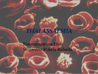
The Thalassaemia Syndromes. Ahmad Sh. Silmi Msc Haematology, FIBMS. The Thalassaemia Syndromes. The thalassaemia are heterogeneous group of inherited disorders, which are characterized by reduced or absent synthesis of one or more globin chain type.

The Thalassaemia Syndromes
E N D
Presentation Transcript
The Thalassaemia Syndromes Ahmad Sh. Silmi Msc Haematology, FIBMS
The Thalassaemia Syndromes • The thalassaemia are heterogeneous group of inherited disorders, which are characterized by reduced or absent synthesis of one or more globin chain type. • The imbalance of globin chain synthesis, which result leads to ineffective erythropoiesis and a shortened red cell lifespan. • In contrast to the structural haemoglobinopathies, the affected globin chain is structurally normal; it is only the rateat which it is synthesized which is affected.
The thalassaemia are most common in part of the world where malaria is, or was recently, endemic: the result of positive selection for a gene, which affords some protection against malaria. The distribution of the different forms of thalassaemia is not uniform: each is most commonly found in certain populations. βThalassaemia is most common in people from the Mediterranean, Africa, India, SE Asia and Indonesia. The incidence of mutations, which lead to βthalassaemia, reaches almost 10% in some parts of Greece. The disorder is relatively rare in Northern and Western Europeans and in native Americans. The clinically mild forms ofαthalassaemia (α +thalassaemia)are most common in American blacks, Indonesia, SE Asia, the Middle East, India, and the Mediterranean. 30% of American blacks are silent carriers of α +heterozygous, while 3% are homozygous. Homozygous express minimal symptoms of disease. The clinically severathalassaemia (α0thalassaemia) are common in people from the Philippines, SE Asia and S China. The population incidence of deletions, which leads to this form, reaches 25% in some parts of Thailand.
Classification The thalassaemias are classified according to three criteria: 1- The affected globin gene(s) e.g. α , β , dδ , etc. 2- Whether the reduction in synthesis in the affected gene is partial (β+) or absolute (β0). 3- The genotype e.g. homozygous β0.
α -Thalassaemia More than 95% of a thalassaemias result from the deletion of one or both of a globin genes located on chromosome 16. This gives rise to five possible genotypes:
Pathophysiology • The myriad manifestation of this complex group of disorders result from the imbalanced synthesis ofα-like and non- α -like globin chains. • Under normal circumstances, the rate of synthesis ofαglobin must be more or less matched by the total synthesis ofβ, δandγ globin chains.
Pathophysiology • Impaired synthesis of αglobin results in the accumulation of unpaired non- αglobins within the developing erythroblasts and vice versa.
Pathophysiology • Unpaired globin chains are unstable: they form aggregates and precipitate within the cell, causing decreased deformability, membrane damage and selective removal of the damaged cell by reticuloendothelial system.
Pathophysiology • Unpaired αglobin chains are extremely insoluble and causes sever damage to the developing erythroblasts. • Unpairedβglobin chains, on the other hand, form haemoglobin H, which is relatively stable and only precipitate as the red cell ages. Thus moderate impairment of βglobin synthesis is associated with a greater degree of ineffective erythropoiesis and haemolysis than an equivalent impairment of αglobin synthesis.
αThalassaemia • The affected individuals in this disease are belonging to one of four groups according to the increasing severity of their symptoms: 1- "silent" carriers 2- αthalassaemia trait 3- haemoglobin H disease 4- haemoglobin Barts hydrops foetalis • The groups correspond approximately to the functional equivalent of thedeletion of 1, 2, 3 or 4aglobin genes respectively.
1- "Silent" carriers • Deletion of a single a globin gene has no significant effect on the affected individual. • As adults, no haematological abnormality can be demonstrated using standard laboratory techniques (excluding DNA analysis). • Umbilical cord blood of newborns may contain 1% of haemoglobin Barts (γ4). • Such individuals can only be defined with complete reliability by DNA analysis.
2- α Thalassaemia Trait • Individuals with deletion of twoαglobin genes may be: • α+ homozygous (α-/α-) or α0 heterozygous (- -/ αα). It's important to know to which group a given individual belong to give accurate genetic counseling. • The two groups are clinically indistinguishable and present identical laboratory results.
Laboratory findings of Thalassaemia Trait Affected individuals typically show: 1- Mild microcytic hypochromic anaemia with no significant symptoms. 2- Precipitated haemoglobin H (- - /α -) can be demonstrated by supravital stain in small minority of red cells. 3- Umbilical cord blood contains up to 10% of haemoglobin Barts.
3- Haemoglobin H Disease • It's arises from the deletion of threeα globin genes. • The severity of Hb H is highly variable. • It's characterized by a moderately sever anaemia and hepatosplenomegally. • Typically, the haemoglobin level is maintained around 8 g/dl, and transfusion support is unnecessary. • Extramedullary haemopoiesis and skeletal abnormalities are uncommon.
Laboratory Findings ThePeripheral blood film includes: • Microcytosis, hypochromasia, fragmented red cells, poikilocytosis, and polychromasia and target cells. • Multiple haemoglobin H inclusions are seen in most of the cells; these bodies cause haemolytic anaemia, which characterizes the condition. • Umbilical cord blood contains up to 40% haemoglobin Barts. • Adult's blood contains between 5-35% of haemoglobin H.
4- Haemoglobin Barts Hydrops Foetalis • The most sever form of a thalassaemia results from the deletion of all four a globin genes and so is associated with a complete absence of a globin synthesis.
4- Haemoglobin Barts Hydrops Foetalis • Because of the absence of a globin synthesis, no functionally normal haemoglobins are formed after the cessation ofζglobin synthesis at about 10 weeks gestation. • Instead, functionally useless tetrameric molecules such as haemoglobin Barts (γ4)and haemoglobin H (β4)are synthesized. • Thus, although the haemoglobin concentration at delivery typically is about 6 g/dl, functional anaemia is much more sever. • The severity of anaemia causes gross oedema secondary to congestive cardiac failure and massive hepatosplenomegally. • Pregnancy usually terminates in a third trimester stillbirth, often after a difficult delivery.
Laboratory Findings • The peripheral blood smear shows marked microcytosis, hypochromasia, poikilocytosis, fragmentation and numerous nucleated red cells. Haemoglobin electrophoresis confirms this abnormality.
β Thalassaemia • βThalassaemia usually results from point mutations within the βglobin gene cluster, βthalassaemia can be classified according to the severity of their symptoms into three groups: 1- βthalassaemia minor (or trait) 2- βthalassaemia major 3- βthalassaemia intermediate
1- β Thalassaemia minor • It's the mildest form, which arises from the inheritance of a single abnormal β globin gene. Typically, the affected individual exhibits no significant signs of the disease, and may be unaware of the condition, and generally live a normal lifespan.
Laboratory findings • Microcytic hypochromic anaemia, with target cells a prominent feature in the peripheral blood film. • Red blood cell count is high to compensate for the generated anaemia. • Haemoglobin level is around 10-11 g/dl. • Reticulocyte is slightly increased. • White blood cells is normal
Bone marrow : Generally shows some degree of erythroid hyperplasia and mild ineffective erythropoiesis. Iron storage is slightly increased. Haemoglobin Electrophoresis: Hb F(2 - 6 % ) Hb A2 ( 3 - 7 %) Hb A (87 - 95 %)
2- β Thalassaemia major • βThalassaemia major results from the inheritance of twobthalassaemia genes. Affected individuals are either homozygous or double heterozygous for two distinct mutations. • In the absence of treatment, the condition is characterized by : 1- sever anaemia 2- gross splenomegally 3- Frequently hepatomegally 4- Retarted growth 5- Facial mongoloid appearance 6- Rarely live beyond the second decay.
Laboratory findings • peripheral blood • Sever haemolytic anaemia with Hb< 7.0 g/dl • Microcytic hypochromic due to decrease globin synthesis. • Marked anisocytosis and poikilocytosis. • Increased polychromatophilia. • Numerous target cells. • Howell-jolly bodies and sedrocyte are common. • Increased NRBC's ( 200 or more / 100 WBC's) • Increased reticulocyte. • WBC is slightly increased with occasional immature granulocyte. • Platelets are slightly increased
2- Bone Marrow: The bone marrow shows erythroid hyperplasia, and excess blood transfusion & haemolysis will lead to precipitation of iron in spleen and liver. 3- Biochemical tests: Haptoglobin is decreased. Bilirubin is increased.
4- Haemoglobin Electrophoresis • Analysis of the haemoglobins present reveals a marked increase in Hb F, the precise value of which is dependent on the genetic defect(s) present. for example: • In homozygous β0 thalassaemia: Hb F accounts for up to 98 % of the total. • In double heterozygous β+ thalassaemia: Hb F accounts for 40-60 % • Hb A2 is increased in both defects. • The increase in d and g chains is a compensatory mechanism due to the decrease in the production of β chain.
Beta thalassemia major treatment • Transfusion • Iron chelation • stem cell transplant
3- β Thalassaemia intermedia Typically, thalassaemia intermedia arise from one of three circumstances: • Inheritance of mildβ thalassaemia mutations. • Co-inheritance of a gene which increases the rate ofγglobin synthesis. • Co-inheritance ofαthalassaemia. Reduction inaglobin synthesis reduces the imbalance in α: non-αglobin synthetic ratio.
3- β Thalassaemia intermedia • Thalassaemia intermedia encompass all cases of β thalassaemia with significant symptoms of disease which do not require regular transfusion to maintain their haemoglobin level above 7 g/dl. • The laboratory and clinical features of this condition mirror those of the more sever phenotype. The major cause of morbidity is due to iron overload as a result of increase gastrointestinal absorption of dietary iron in anaemic patients; these results in increase total body iron. • The bone marrow is massively imposed by erythroid hyperplasia, this leads to increase demand of iron, which exceeds the supply capacity of the reticuloendothelial system. Thus functional iron deficiency is present, despite raised in iron stores.
What Is Thalassemia? • Thalassemia is an inherited blood disorder that causes mild or severe anemia (uh-NEE-me-uh). The anemia is due to reduced hemoglobin (HEE-muh-glow-bin) and fewer red blood cells than normal. Hemoglobin is the protein in red blood cells that carries oxygen to all parts of the body.
In people with thalassemia, the genes that code for hemoglobin are missing or variant (different than the normal genes). Severe forms of thalassemia are usually diagnosed in early childhood and are lifelong conditions.
The two main types of thalassemia • alpha and beta, are named for the two protein chains that make up normal hemoglobin. The genes for each type of thalassemia are passed from parents to their children. Alpha and beta thalassemias have both mild and severe forms.
Alpha thalassemia • occurs when one or more of the four genes needed for making the alpha globin chain of hemoglobin are variant or missing. Moderate to severe anemia results when more than two genes are affected. The most severe form of alpha thalassemia is known as alpha thalassemia major. It can result in miscarriage.
Beta thalassemia • occurs when one or both of the two genes needed for making the beta globin chain of hemoglobin are variant. The severity of illness depends on whether one or both genes are affected and the nature of the abnormality. If both genes are affected, anemia can range from moderate to severe. The severe form of beta thalassemia is also known as Cooley’s anemia. Cooley’s anemia is the most common severe form of thalassemia in the United States.
Alpha Thalassemias • Alpha thalassemia “silent carrier” • Mild alpha thalassemia, also called alpha thalassemia minor or alpha thalassemia trait • Hemoglobin H disease • Hydrops fetalis, or alpha thalassemia major
Beta Thalassemias • Beta thalassemia minor, also called thalassemia minor or thalassemia trait • Beta thalassemia intermedia, also called thalassemia intermedia or mild Cooley’s anemia • Beta thalassemia major, also called thalassemia major or Cooley’s anemia • Mediterranean anemia
Cooley ’s anemia • Cooley’s anemia is another name for the severe form of beta thalassemia. The name is sometimes used to refer to any type of thalassemia that requires treatment with regular blood transfusions.
Thalassemia is caused by variant or missing genes that affect how the body makes hemoglobin. Hemoglobin is the protein in red blood cells that carries oxygen. People with thalassemia make less hemoglobin and fewer circulating red blood cells than normal. The result is mild or severe anemia
Many possible combinations of variant genes cause the various types of thalassemia. Thalassemia is always inherited (passed from parents to children). People with moderate to severe forms of thalassemia received variant genes from both parents. A person who inherits a thalassemia gene or genes from one parent and normal genes from the other parent is a carrier (thalassemia trait). Carriers often have no signs of illness other than mild anemia, but they can pass the variant genes on to their children.
Hemoglobin includes two kinds of protein chains called alpha globin chains and beta globin chains. If the problem is with the alpha globin part of hemoglobin, the disorder is alpha thalassemia. If the problem is with the beta globin part, it is called beta thalassemia. There are both mild and severe forms of alpha and beta thalassemia. Severe beta thalassemia is often called Cooley’s anemia.
Alpha Thalassemia Four genes are involved in making the alpha globin part of hemoglobin—two from each parent. Alpha thalassemia occurs when one or more of these genes is variant or missing.
People with only one gene affected are called silent carriers and have no sign of illness. People with two genes affected (called alpha thalassemia trait, or alpha thalassemia minor) have mild anemia and are considered carriers. People with three genes affected have moderate to severe anemia, or hemoglobin H disease. Babies with all four genes affected (a condition called alpha thalassemia major, or hydrops fetalis) usually die before or shortly after birth.
If two people with alpha thalassemia trait (carriers) have a child, the baby could have a mild or severe form of alpha thalassemia or could be healthy.
Beta Thalassemia Two genes are involved in making the beta globin part of hemoglobin—one from each parent. Beta thalassemia occurs when one or both of the two genes are variant.