Download
1 / 36
380 likes | 970 Views
Arrhythmias and Antiarrhythmic Drugs. Class I (Na + channel blockers) 1A: procainamide, quinidine (no longer recommended) 1B: lidocaine 1C: flecainide, propafenone Class II ( -blockers) non-selective: propranolol selective: metoprolol Class III (K + channel blockers)

E N D
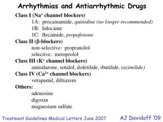
Arrhythmias and Antiarrhythmic Drugs Class I (Na+ channel blockers) 1A: procainamide, quinidine (no longer recommended) 1B: lidocaine 1C: flecainide, propafenone Class II (-blockers) non-selective: propranolol selective: metoprolol Class III (K+ channel blockers) amiodarone, sotalol, dofetilide, ibutilide, (azimilide) Class IV (Ca2+ channel blockers) verapamil, diltiazem Others: adenosine digoxin magnesium sulfate AJ Davidoff ‘09 Treatment Guidelines Medical Letters June 2007
What happens to PR interval if AV nodal conduction is prolonged? What happens to QRS interval if conduction through heart is slowed? What happens to QT interval if APD is prolonged?
ACh M2 ACh receptors IK+ , ICa2+, If NE 1-AR If ICa2+ (L-type) Nodal cell firing rate control ACh = acetylcholine M-ACh = muscarinic ACh NE = norepinephrine -AR = adrenergic receptor I = whole cell current Sherwood Fig 9-24
ITO Fast Action Potentials IKr Outward K+ currents (delayed rectifiers) Fast Na+ current (INa) IKs ITO = transient outward K+ current
Optional information inward positive current NCX = sodium/calcium exchanger outward positive current IKACh = ACh K+ current ICaL = L type Ca2+ current INa = Na+ current Purkinje fiber transient outward K+ current ultra rapid K+ current current activation rapid K+ current slow K+ current ‘funny’ current Nattel and Carlsson 2006 Nature Reviews; Drug Discovery 5:1034-1049
Most arrhythmias result from altered conduction and/or automaticity Conduction abnormalities Typically arise from partial depolarization due to injury (e.g., over stretch, ischemia) or abnormal anatomy Partial or complete block Accessory conduction pathways e.g., Wolff-Parkinson-White (WPW) Syndrome Re-entry Fibrillation (multi-re-entry loops)
Automaticity abnormalities • Originating from nodal cells or ectopic loci • Depolarization-dependent automaticity: • Changes in sinus node firing rate • Prolonged action potential duration (APD) • Early afterdepolarizations • may lead to premature ventricular contractions (PVCs) or multiple extrasystoles • Long QT syndrome • may lead to Torsades de Pointes
In terms of cellular target and action potential (AP) duration, what strategy would you use for: Rapid nodal firing? Supraventricular tachycardias? Premature ventricular contractions (PVCs)? Ectopic ventricular arrhythmias? Ventricular tachycardias? • Slow SA or AV nodal depolarizations • Slow atrial cell conduction • Slow AV conduction • Slow ventricular conduction • Prolong ventricular AP duration • Shorten ventricular AP duration
Strategies to convert fibrillation/tachycardia For acute atrial fibrillation or supraventricular tach. Target atrial muscle cells or AV nodal tissue hyperpolarize membrane conduction velocity AV node Drugs adenosine Ca2+ channels -blockers digoxin (nodal cell)
For V. fib. or V. tach. Target ventricular muscle cells • conduction velocity (Vmax) • e.g., block Na+ channels (Class I drugs) AP duration (APD) (refractory period) e.g., block K+ channels (now typically preferred over Class I drugs)
For Maintenance (preventing re-occurrence) • Slow AV nodal conduction or frequency of firing • Drugs: digoxin • Ca2+ channel blockers • -blockers • Na+ channel blockers are contraindicated for: • long-term therapy • patients with structural defects (e.g., fibrosis, WPW)
Cellular models of arrhythmias Increased automaticity: sympathetic activity (e.g., NE or Epi) vagal activity (e.g., drug-induced, quinidine) TP = threshold potential MDP = maximum diastolic potential Brenner Box 14-1
normal -40mV -60mV -90mV Ectopic pacemaker activity Often due to partial ischemia, resulting in a more postive resting membrane potential Na+ channels inactivated Vmax Conduction
Closed (ready to open) Resting potential (-90 mV) Open Threshold and activation potentials (-50 mV to +30mV) Closed (unable to open) Inactivation potentials (+30 mV to -90mV) see also Katzung Fig 14-2 Sherwood Fig 4-7
Katzung Fig 14-8 Abnormal Impulse Conduction (hypothetical model) Normal conduction Ischemia Re-entry loop Unidirectional block
Abnormal Impulse Conduction • Ischemic or fibrotic areas slow conduction • Ischemia partially depolarizes resting membrane potential, inactivates some Na+ channels • Slow rate of phase 0 (i.e., rapid depolarization phase) results in slow conduction through heart Re-entry loops A model for unidirectional block Boron Fig. 20-14
Afterdepolarizations (due to abnormal intracellular Ca2+ regulation) ‘Delayed’ EADs DADs EADs prolonged APD Clinical arrhythmia: e.g., torsades de pointes due to: long QT syndrome genetic defects (HERG) disease drug-induced DADs HR or [Ca2+]i Clinical arrhythmia: e.g., Ca2+ overload due to: digoxin or phosphodiesterase (PDE) inhibitor toxicity Brenner Box 14-1
Boron Fig. 20-15 If afterdepolarization is large can trigger PVC If sustained, can trigger “run” of extra systoles
Nature of Antiarrhythmic Drugs • All have potential of being pro-arrhythmic: • Toxicity may depress automaticity or • depress conduction velocity • Many are metabolized by cytochrome P450 enzymes • (induced/inhibited, “poor metabolizers”) • Most have a low TI • (especially Na+ channel blockers) • Most show ‘use- (or frequency-) dependent block’ • higher affinity for membranes depolarizing frequently • Advantage, because drugs may be selective for abnormally fast rhythms Generally classified based on primary mechanism of action
Class Phase O Depression Repolarization Action Potential Duration IA Moderate Prolonged Increased IB Weak Shortened Decreased IC Strong No effect No effect Class I Na+ channel blockers Na+ channels inactivated “use-dependent block” resting/closed Brody Table 14-3
What might happen? Class 1A Block Na+ channels and K+ channels • No longer drugs of choice • Indications: (alternative DOC) • Atrial fibrillation or flutter • SVT • Ventricular fib or tachycardia Vmax APD • Toxicity includes: • Prolongs APD too much • Antimuscarinic effects • (may inhibit vagus n.) Quinidine (oral) prototype Class IA rarely used anymore Procainamide (oral or IV) less (-) on vagus Brenner Fig 4-2
Class 1B Block inactivated Na+ channels Rapidly binds to depolarized membranes (e.g., during ischemia) Rapidly dissociates from resting cells • Indications: • Ventricular tachycardia • V. re-entrant loops? (PVCs) • during surgery • No effect on atrial cells • (with short APD) Vmax APD • Toxicity: • Relatively safe (hemodynamically) • but efficacy is relatively low Lidocaine (IV only)
Class 1C Block open, closed and inactivated Na+ channels Very slow off rates, not selective for fast rhythms Indications: (alternative drug of choice) Sustained ventricular tachycardia Paroxysmal A. fib or SVT only with no signs of structural heart disease (e.g., ischemia, hypertrophy) Vmax • APD • Toxicity: • Slows conduction (Vmax) too much • Can cause re-entrant loops • (especially v. arrhythmias) Flecainide Propafenone (also ~-blocker) Flecainide mortality after acute MI (CAST; cardiac arrhythmia suppression trial)
Class II (-blockers) Block -AR on nodal and muscle cells: HR A-V conduction (may contractility) Slow rate of depolarization of phase 4 (pacemaker potential) Indicated for: Acute/chronic A. Fib and Flutter Long term SVT IV or PO Brenner Fig 4-4 Propranolol (non-selective) Metoprolol (1 selective) Some may be cardioprotective after acute MI
Class IV (Ca2+ channel blockers: cardioselective) • Inhibit L-type Ca2+ channels • Effectively raise threshold potential to fire an AP • Use-dependent block, therefore more effective with fast HR • HR, A-V conduction velocity, (may contractility) Indicated for: Acute/chronic A. Fib and Flutter Acute/chronic SVT Verapamil (more effect on A-V conduction) Diltiazem (more effect on SA nodal cells) Dihydropyridines (DHPs) have little antiarrhythmic activity
Class III (K+ channel blockers) Block delayed rectifier channel (IKr) (as well as other channels) Indicated for SVT, A. fib, V. fib and V. tach APD Amiodarone (DOC) also Na+, Ca2+ channel blocker and -blocker Sotalol also -blocker (non-selective) Pure Class III blockers Dofetilide (PO only) Ibutilide (IV only) Azimilide(blocks IKr and IKs) risk of torsades de pointes (not with amiodarone)
Others Antiarrhythmic Drugs Digoxin Inhibits Na/K ATPase Slows A-V conduction (through increasing vagal tone) Increases refractory period Indicated for: A. Fib with fast ventricular rate* (and CHF) Toxicity: complete heart block (narrow TI) may precipitate Ca2+ overload (e.g., torsades) *approaches are now focusing on controling heart rate (with warfarin), rather than rhythm (G. Wyse, AHA website updated 5/08). Thus digoxin is used much less frequently now.
Digoxin: Cardiac effects: Increases intracellular [Na+], increases in Ca2+ (via NCX) more Ca2+ to trigger SR Ca2+ release, increases contraction (positive inotropic effects, discussed in heart failure lecture) Decreases intracellular [K+], depolarizes membrane potential partially inactivates Na+ channels in fast fibers, reduces excitability, slows conduction High affinity to vagus nerve (particularly at the AV node), increases vagal tone slows AV nodal conduction Binds to, and inhibits Na+/K+ ATPase pumps in other tissues (non-cardiac toxicities include visual distrubances -yellow hues), with highest affinity to cardiac and vagal nerve.
Adenosine Opens K+ channels hyperpolarizes membrane (also blocks Ca2+ channels) Selective for coronary arteries and atrial muscle cells (not ventricular myocytes) Slows SA nodal firing Slows A-V conduction Very short T1/2 (seconds) Indicated for ‘cardioconversion’ • Magnesium • Inhibits Ca2+ influx through L-type Ca2+ channels • Indicated for: • Drug-induced torsades • Digoxin-induced ventricular arrhythmias
Triggered activity due too much intracellular Ca2+ [Ca2+]i Na/Ca exchange [Na+]i (3Na+(in): 1Ca2+(out)) (depolarize membrane) Mg2+ (for torsades) Open L-type Ca2+ channels
Effects of serum potassium appear paradoxical: contrary to what would be predicted by changes in electrochemical gradient • Hyperkalemia • Reduces action potential duration (APD) • Slows conduction • Decreases pacemaker rate and arrhythmogenesis • (leading to bradiacardia and perhaps asytole) Hypokalemia (more detrimental than hyperkalemia) • Prolongs APD • Increases pacemaker rate and arrhythmogenesis • (increasing risk of ventricular fibrillations) • Increases sensitivity to K+ channel blockers • resulting in accentuated APD prolongation with risk of Torsades de Pointes Katzung 2009 p. 228 Ranolazine Recently approved for chronic stable angina Prolongs QT interval (maybe by inhibiting late Na+ current or delayed K+ rectifier current)
In terms of cellular target and action potential duration, what strategy would you use for: Rapid nodal firing? Supraventricular tachycardias? Premature ventricular contractions (PVCs)? Ventricular tachycardias? Na+ channel blockers -blockers K+ channel blockers Ca2+ channel blockers
Atrial fibrillation or flutter: • Acute • Rate control: (IV) verapamil, diltiazem, -blockers, digoxin • Chronic • Rate control: verapamil, diltiazem, -blockers, digoxin • Maintenance of sinus rhythm: • amiodarone, sotalol, flecainide, propafenone, dofetilide • Other SVTs: • Acute • (vagotonic maneuvers, e.g., carotid sinus massage) • (IV) adenosine, verapamil, diltiazem • Chronic • -blockers, verapamil, diltiazem, flecainide, propafenone, amirodarone, sotalol, digoxin According to Treatment Guidelines, Medical Letters 2007
PVCs or non-sustained V. tach: • Asymptomatic • no therapy • Symptomatic(flecainide is contraindicated post-MI) • -blockers (post MI improves mortality rates) • Sustained V. tach. or V. fib: • Acute • DC cardioversion is safest • amiodarone • Chronic • Implantable cardiac defribillator (ICD) (NEJM Jan 20, 2005) • amiodarone, plus a -blocker According to Medical Letters
Alternative Classification based on target Drug therapy for supraventricular arrhythmias Adenosine (IV only) Verapamil Diltiazem Esmolol (IV only) Ibutilide (IV only) Dofetilide (oral only) Drug therapy for ventricular arrhythmias Procainamide (not preferred) Lidocaine (IV only) Flecainide or Propafenone (oral, not approved for IV in US) Sotalol Amiodarone LH Opie 2004