Download
1 / 144
2.92k likes | 6.42k Views
Physical Therapy Examination. PTP 565: Fundamentals of Tests and Measures. Physical Therapy Examination.

E N D
Physical Therapy Examination PTP 565: Fundamentals of Tests and Measures
Physical Therapy Examination • Examination: gathering of data: history, systems review, selecting and administering tests and measures. Comprehensive screening and specific testing process. Leads to a diagnostic classification. • APTA Guide to Physical Therapy Practice
PHYSICAL THERAPY EXAMINATION • Important to develop a system for examination
Organizational Structure of an Orthopedic Exam • History • Systems Review • Scanning Exam
IV. Tests and Measures • Observation/Posture/Gait • Active ROM • Passive ROM • Resisted movements • Neuro Screen • Functional Assessment • Special Tests • Joint Play • Palpation
I. History Benefits from obtaining a history: 1. report from the patient of their condition 2. assists in formulating a working hypothesis for differential diagnosis 3. gives clinical signs and symptoms 4. assists with formulating an examination plan. 5. assists in setting functional goals for the patient
General Demographics • Gender/Age/Height/Weight • Social History • Occupation, Employment, Work • Functional Status, Activity Level • Growth and Development • Living Environment • Red Flags
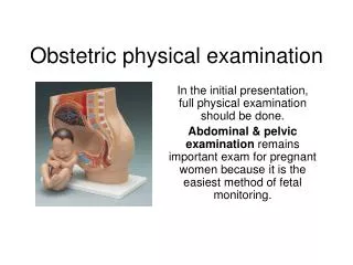
Other Significant Information • Past Medical History • Past Surgical History • Medication • Review of Systems: Questionnaire information about Heart, Lungs, Other organs.
Red Flags • Definition: • Constitutional Signs and Symptoms • Fevers, chills, night sweats • Malaise or Fatigue • Unexplained Nausea or vomiting • Recent unexplained weight changes • Unilateral, bilateral or quadrilateral paresis or paresthesia • Shortness of breath • Bowel or bladder dysfunction
Red Flags: continued • Insidious onset of pain • Multiple levels of neurological symptoms • Pain at night • Increase in pain intensity over time
Red Flags: • No Red Flags? Continue with the Exam • Red Flags exist: Clinical Decision • What do you do with the patient? • Refer • Consult • Send to the ER • Call an ambulance • Make an appointment for the Primary Physician
Onset Sudden After 24 hours Gradual or insidious onset Intensity Visual Analog Scale Verbal Analog Scale McGill-Melzack pain questionnaire Thermometer Pain Rating Scale Pain Perception Acute Subacute Chronic Pain Progression Better Worse No Difference Pain
Quality of Symptoms Location of Symptoms Aggravating Factors Easing Factors Pain Descriptors Sharp Shooting pain Aching Burning Pins and Needles Numbness Stiffness Twinges
How Acute is the injury? • In terms of pain • Time since onset of symptoms • Length of Time pain lasts • Acute • Sub-acute • Chronic • Other Symptoms
Signs/symptoms of acute conditions • Inability to maintain any position for any length of time. • Verbal and nonverbal expressions of pain while moving, slow guarded movements, • Pain Scale that is very high 9-10 on a scale of 1-10 • Recent injury like within 1-2 days
Examination for acute conditions: Key Points Don’t try and complete the examination Move into an intervention designed to relieve acute symptoms Re-examine at a later date when the individual is able to tolerate the activity Goal: interrupt the pain cycle
Injury not in acute phase: • Continue with the examination process.
II. Systems Review • Cardiovascular System • Integumentary System • Musculoskeletal System • Neuromuscular System • Psychological Status
III. Screening Examination • Purpose: decide area of involvement so that the examination can be directed to the appropriate area
Scanning exam is especially helpful when: • No history of trauma • Radicular signs are present: referred pain along a specific spinal nerve root distribution (dermatome) • History of trauma along with radicular signs are present • Patient reports pins and needles sensation (paraesthesia) or complete numbness (anesthesia) in the region.
“long track” signs (spinal cord or UMN lesions) are present (flaccidity or spasticity) • patients signs and symptoms don’t reveal any clear pattern • ? psychological factors, secondary gain or patient is a poor historian
Scanning Exam: Upper Quadrant • Upper Quadrant: areas to scan include the joints above or below the patient’s dysfunction. • Cervical spine: must screen the thoracic spine, TMJ, and shoulder • Shoulder: must screen the cervical spine, thoracic spine, elbow and hand • Elbow: must screen the shoulder, thoracic spine, hand • Hand: must screen shoulder and elbow
Thoracic Spine Scan • Thoracic Spine: in particular must be aware of the systemic dysfunctions which show up as thoracic pain, rib pain, and or/shoulder pain • Upper: cervical spine, shoulder, ribs • Lower: lumbar spine, ribs
Lower Quadrant: • Lumbar spine: must screen the thoracic spine, ribs, sacroiliac joint, hip, sometimes knee and feet • Sacroiliac joint: lumbar spine, hip, knee, ankle • Hip: lumbar spine, SI, knee, ankle • Knee: lumbar spine, hip, ankle • Ankle/Foot: lumbar spine, knee
What do you check during a scanning exam? • Observation/Posture • Active ROM • Passive ROM with Overpressure • Muscle Strength • Sensory: myotome, dermatome and reflexes
After the Screening Exam Clinical Decision Keep ReferKeep and Refer (Consult)
Keep/Refer/Consult • Keep: Patient is appropriate for Physical Therapy • Refer: Patient’s signs and symptoms do not have a musculoskeletal origin • Consult: Patient can be treated by physical therapy but also needs another medical professional for assistance
Specific Joint Examination • Investigative hat • Ruling out or confirming hypothesis for the dysfunction • Determining the facts: muscle weakness, loss of range etc.
Observation: • Done before any hands on activities. • Looking phase of exam • Observe/Inspect for Visible defects, dysfunctions, alignment abnormalities
Observation • Gait: quick observation noted while the patient is walking from the waiting room to the examination room. • Will note gait deviations that are really obvious like an antalgic pattern. • Can also get an idea of balance by looking at base of support, position of arms, waddling gait • Assistive devices used by patient
Transitional movements • Sit to stand, walking, back to sitting • Guarded movements, stiffness, willingness to move • Ability to maintain a position like sitting or standing
Facial expression during history taking, affect of patient – anxious, flat etc. • Patient’s willingness to move, substitute movements noted • Medicated? Too much or too little?
Posture: review standard alignment; sagittal and A/P views, look at symmetry and determine if the findings are related to the dysfunction.
UE: head, shoulder position in relation to acromian, thoracic spine position, Q angle of elbows, previous fractures for example a clavicular fracture,
Obvious deformities are noted during the posture exam such as scoliosis, or presence of bony abnormalities in the LE • LE – may observe patella positioning in relation to the LE alignment, Varus or valgus position of knees, genu recurvatum • Ankle position
Other Observations: • Muscle wasting • Scars and color of scars. If red these are recent, pale, they are old and well healed. • Note any joint noise such as crepitus, snapping or grinding sounds made with movement • Skin changes, nail changes can be observed http://www.cdaarthritis.com/images_slides/images_slides_29e_larger_slides.htm
Purpose of HANDS ON examination: • To rule out or confirm a hypothesis developed by the therapist during the history and observation as to the patient’s cause of pain.
Active ROM • AROM • Patient’s willingness to move • Joint mobility/limitations • Muscle control • Muscle power • Movements which cause pain
AROM: What to observe • When/where pain occurs during movement • Does the movement increase the intensity of the pain • Reaction of the patient to pain • Amount of observable restriction/limitation • Pattern of movement • Willingness of the patient to move the limb • Quality of movement
2. Passive movement: • This is done with overpressure after the patient has moved through their available range. • Determines end feel at that particular joint with that physiological movement. • End feel: quality of resistance to movement at the end of a physiological movement
Normal end feels • Hard: Bone to bone • Firm: Capsular end-feel, Tissue Stretch • Soft: Soft tissue approximation
Abnormal end feels • Muscle spasm: Sudden and abrupt stop to movement • Capsular: similar to tissue stretch, it doesn’t occur at end range • Bone to bone: similar to the normal bony end feel but the restriction in range occurs earlier than it should in the normal range. • Empty: pain is produced with movement • Springy Block: elastic resistance to further movement
Definitions: • Gonia: Angle • Metron: Measure • Definition: Measurement of the angles created by rotary motion of two bones around a joint
Goniometry Purposes: • Joint Position • ROM • Active ROM • Passive ROM
Other Aspects 1. Only motions occurring in a single plane are measured with few exceptions. 2. Measures osteokinematic motion NOT arthrokinematic motion 3. Flexibility Measurement
4. Degrees of freedom: freedom of movement around a plane of motion • Motion occurs around one plane – one degree of freedom • Motion occurs around two planes – two degrees of freedom • Motion occurs around three planes – three degrees of freedom
Purposes of Joint ROM Examination 1. Database: • Establish the existing ROM available in the joint • Compare this with the normal ranges given in the literature • Compare with the other side. • Beginning data base for treatment
2. Aids in diagnosing and determining joint function a. Hypomobility: less than normal ROM when compared to established norms or the patients other side. b. Hypermobility: greater than normal ROM when compared to established norms or the patients other side.
C. Capsular Pattern: • Cyriax: pattern of limitation in range of motion at a joint which predictably occurs when the capsule of the joint is tight due to capsular fibrosis or extended due to swelling. • Fixed proportional loss of range of motion, it is not all inclusive as he never considered flex/extension when determining the shoulder’s capsular pattern.
D. Orthotic fabrication: Many splints and orthotics rely on goniometric measurement to fabricate the devices