GU IMAGING 2012
GU IMAGING 2012. The kidneys and adrenal glands are retroperitoneal structures. The kidneys have direct blood flow from the aorta and to the IVC. TAKE HOME POINTS. 1- OBSTRUCTION VS NON OBSTRUCTION 2 - SOLID VS CYSTIC 3- PREGNANT OR NOT 4- INTRAUTERINE OR ECTOPIC. ANATOMY.

GU IMAGING 2012
E N D
Presentation Transcript
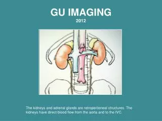
GU IMAGING2012 The kidneys and adrenal glands are retroperitoneal structures. The kidneys have direct blood flow from the aorta and to the IVC.
TAKE HOME POINTS 1- OBSTRUCTION VS NON OBSTRUCTION 2 - SOLID VS CYSTIC 3- PREGNANT OR NOT 4- INTRAUTERINE OR ECTOPIC
ANATOMY • KUB-plain x-ray • IVP • CT • NUCLEAR MEDICINE • ULTRASOUNDand MRI • ANGIOGRAPHY Ultrasound and CT are used as first line tools in GU disease imaging.
LT. KIDNEY RT. KIDNEY PSOAS MUSCLE PSOAS MUSCLE PSOAS MUSCLE R The kidneys are not always seen on an abdomen x-ray but can sometimes be outlined by retroperitoneal fat. The psoas muscle may be seen for a similar reason.
THE BLADDER IS VISUALIZED BECAUSE IT IS OUTLINED BY FAT—A LOWER DENSITY.
INTRAVENOUS PYELOGRAM – IVP INTRAVENOUS IODINE CONTRAST WITHOUT CONTRAST-plain or scout film ARTERIOGRAM INTRAARTERIAL IODINE CONTRAST 6
IVP INTRAVENOUS PYELOGRAM KUB KIDNEY- URETERS- BLADDER R R The administration of iodinated IV contrast can image the GU tract with radiographs---IVP. This exam is not done as frequently now as in past and more imaging is by ultrasound and CT.
PELVIS MALE FEMALE CONTRAST IN DISTAL URETER BLADDER PROSTATE UTERUS The prostate gland impresses the bladder normally in males but enlarges with prostatic hypertrophy. BLADDER Note superior impression on bladder due to uterus.
IVP Female abdomen SI JOINT BODY OF UTERUS Note superior impression on bladder due to uterus. BLADDER
CT SCANSwithout and with contrast administration The IV contrast which is being filtered and increasing density of the renal tissue. Compare with liver density.
A 65-year old man needs an emergency CT scan with IV contrast. Which of the following is a risk factor for development of contrast induced acute tubular necrosis? • Hypertension • Pheochromocytoma. • Urinary tract infection • Diabetes • Immune-compromised state 10 Countdown
NUCLEAR MEDICINE RENOGRAM This study measures radioactivity as it moves through the kidneys over time. Regions of interest are drawn on the computer and counts are calculated for 30 minutes. This generates functional imaging with data indicating peak time, rate of excretion and percent function The IV administration of the radiopharmaceutical allows for sequential imaging to assess renal function. This is a pharmaceutical agent which is secreted by the kidneys and has a radioactive tag allowing imaging and quantification.
ULTRASOUND LIVER MORRISON’S POUCH ( A POTENTIAL SPACE) Longitudinal Scan Of The Right Kidney Image of Rt. kidney, liver and Morrison's pouch. The echogenic signal in the center of the kidney is renal sinus fat.
THE VALUE OF ULTRASOUND IN INITIAL EVALUATION OF RENAL FAILURE IS: 10 • Low cost • Portability • Lack of radiation • Evaluating blood flow • Excluding obstruction Countdown
ABDOMINAL ARTERIOGRAM CATHETER RENAL ARTERIOGRAM Here arterial contrast is injected into the aorta and immediate imaging shows the arterial lumen outlined by the iodine.
MRARTERIOGRAM Gadolinium Injection Standard Renal Arteriogram Catheter Injection Of Iodinated Contrast Venous injection of gadolinium contrast at MR can outline the arterial tree without the risk of catheterization. The detail is not as good as arterial imaging but is a lower risk procedure.
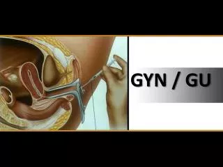
HYSTEROSALPINGOGRAM Contrast has been injected through a cannula placed into the cervical os. Iodinated contrast flows retrograde with injection filling the uterine cavity with reflux into the fallopian tubes. UTERINE CAVITY FALLOPIAN TUBE FIMBRIAE FREE CONTRAST IN PERITONEAL CAVITY This is used to assess infertility due to tubal obstruction. CANNULA
PELVISTrans-abdominal Scanning BLADDER UTERUS The uterus is imaged abdominally using the bladder as an acoustic window to displace bowel and allow visualization.
ULTRASOUND - SAGITTAL Trans vaginal Scanning Endometrium Better detail of the endometrium is shown using the transvaginal ultrasound probe. A higher megahertz transducer with better resolution but less depth penetration is used.
TESTICULAR ULTRASOUND Used to assess for masses and blood flow. Flow can be assessed by Doppler Small parts scanning, testicular, thyroid and carotid uses high megahertz transducers for better resolution.
PATHOLOGY • Congenital • Trauma • Infection • Vascular • Malignant
CONGENITAL ABNORMALITYHorseshoe Kidney KIDNEY AORTA Ascent stopped by Inferior Mesenteric Artery
CROSSED FUSED KIDNEY Pelvic Kidney Embryologically the kidneys can be fused on one side or be ectopic -- ie. Pelvic location
ADULT POLYCYSTIC DISEASEHepatic and Renal Multiple cysts are present in kidneys leading to renal failure in adulthood. Hepatic and Pancreatic cysts can coexist. Increased risk of cerebral aneurysm
Bicornuate uterus is a abnormality of Mullerian duct development and can hinder pregnancy. Septate Uterus Normal
BENIGN RENAL CYST Fluid filled benign renal cyst shows fluid density at CT and lack of echoes at Ultrasound.
WHICH OF THE FOLLOWING IS TRUE REGARDING RENAL INJURY IN TRAUMA? In penetrating trauma absence of hematuria rules out renal injury. In blunt trauma, microscopic hematuria alone is rarely associated with renal injury. Most renal injuries require operative repair. Renal injuries are extremely uncommon in children with blunt trauma Plain radiography is the imaging test of choice for diagnosis. 10 Countdown
TRAUMA Fractured Kidney Horse stepped on patient
VESICO URETERAL REFLUX catheter Reflux from filling bladder with contrast. This can lead to pyelonephritis and scarring . Moderate and severe examples
WHICH OF THE FOLLOWING IS TRUE REGARDING DIAGNOSIS OF KIDNEY STONES? 1-Normal urinalysis essentially rules out the diagnosis. 2-KUB radiograph has ˃90% specificity. 3-Most kidney stones are radiolucent. 4-Ultrasonography has ˃90% sensitivity. 5-CT scan has roughly 90% sensitivity and specificity. 10 Countdown
RENAL CALCULI CT KUB
RENAL ULTRASOUND Sagittal Scan of Rt. Kidney Stone Echogenic stone shows shadowing
ULTRASOUND Kidney Hydronephrosis Dilated collecting system is shown as enlarged fluid filled system on ultrasound. Resolution
Hydronephrosis Stone Note left sided dilated collecting system Stone
A 50-year-old mad develops acute onset of severe right flank pain. A CT scan demonstrates a passed kidney stone in the bladder. The patient has never had a kidney stone before. He asks you what his risk of getting another stone is. You tell him that the lifetime risk of recurrence is approximately: • <1% • 10% • 25% • 50% • 75% 10 Countdown
RENAL ARTERY STENOSIS Renal angioplasty can restore normal perfusion. This can treat hypertension caused by Renin-Angiotensin effect of hypo perfused kidney. RENAL ARTERY STENOSIS CATHETER Delayed function on affected side
DECREASE IN BLOOD PRESSURE RENIN/ANGIOTENSEN CASCADE ANGIOTENSION I---II VIA ANGIOTENSIN CONVERTING ENZYME (ACE) ANGIOTENSIN II CONSTRICTS EFFERENT ARTERIOLE MAINTAINING FILTRATION AT GLOMERULUS
GLOMERULUS Afferent arteriole Efferent arteriole CAPTOPRIL (ACE INHIBITOR)—SITE OF ACTION
RENAL ARTERY STENOSIS Renal angioplasty can restore normal perfusion. This can treat hypertension caused by Renin-Angiotensin effect of hypo perfused kidney. BALLOON RENAL ARTERY STENOSIS CATHETER Dilatation to treat focal renal artery stenosis with catheter and stent placement to maintain vessel size.
RENAL ARTERY STENT • Stents are easily seen on • plain radiographs.
RENAL MALIGNANCY Ultrasound and CT Solid lesion on ultrasound and soft tissue mass on CT is most suspicious for Renal cell carcinoma.
TRANSITIONAL CELL CARCINOMA normal Transitional cell carcinoma is a urothelial lesion shown as filling defect in calyces, ureter or bladder.
Pediatric malignancy Wilm’s tumor and Neuroblastoma Intra renal Supra adrenal Lowe L H et al. Radiographics 2000;20:1585-1603
Neuroblastoma compared to Wilms tumor... 10 1. Metastasizes to lungs more 2. Area more bilateral 3. Less likely Calcify 4. Can cause hypertension 5. Can be in atypical locations more often Countdown
SOLID HYPOECHOIC TESTICULAR LESION Normal Solid lesions in testes are suspicious for malignancy and usually need biopsy.
TESTICULAR TORSION Twisting of testes (torsion) can obstruct blood flow and infarct the testes. Note normal Doppler flow in the left and absent flow on the right.