NOSTRIL
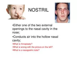
NOSTRIL. Either one of the two external openings to the nasal cavity in the nose; Conducts air into the hollow nasal cavity; What is rhinoplasty? What is wrong with the picture on the left? What is a nasogastric tube?. TURBINATES.

NOSTRIL
E N D
Presentation Transcript
NOSTRIL Either one of the two external openings to the nasal cavity in the nose; Conducts air into the hollow nasal cavity; What is rhinoplasty? What is wrong with the picture on the left? What is a nasogastric tube?
TURBINATES • Thin curved bones along the wall of the nasal passage that increase surface area. • Structure inside the nose covered with a thin membrane that secrete mucous that moistens and filters air. • The turbinates are lined with capillaries that warm and humidify air.
TURBINATES • What is the purpose of the turbinates?
PHARYNX • NASOPHARYNX: cavity forming the upper part of the pharynx at the back of the throat extending from the nasal cavity • OROPHARYNX: cavity forming the upper part of the pharynx at the back of the throat extending from mouth • HYPOPHARYNX: the hypopharynx (or laryngopharynx) is the bottom part of the pharynx, and is the part of the throat that connects to the esophagus • Collectively the pharynx is the intersection between the trachea and the esophagus.
PHARYNX • LARYNGOPHARYX is also known as the HYPOPHARYX.
GLOTTIS • The opening of the trachea
EPIGLOTTIS • The glottal opening is protected by a flap like structure (epiglottis) that protects food from entering the trachea.
LARYNX • Also known as the “voice box”, houses the vocal cords which are 2 folded structures. • When you breathe, there is a large gap between the 2 cords. When you speak, muscles contract bringing the cords closer. The air that passes through the vocal cords vibrates the cords producing sound.
TRACHEA • Also known as the “windpipe”. • Flexible tube that is the passageway for air. It is supported by semi-circular cartilaginous rings. This ensures it does not collapse or interfere with the passage of food in the esophagus. It is lined with ciliated cells that secrete mucous.
TRACHEA • What is a tracheotomy? • What purpose does it serve?
Heimlich maneuver • What is the purpose of this maneuver?
CILIATED CELLS • Work with goblet cells that secrete mucous. • The mucous traps foreign material (dust, bacteria) and the movement of the cilia propel the material back to the nose and throat where it can be expelled by coughing or sneezing.
CILIATED EPITHELIUM • What are goblet cells? • What purpose does the ciliated epithelium provide?
BRONCHI • The trachea ends by branching into 2 bronchi (left and right bronchus). • Lined with ciliated mucous membrane.
BRONCHIOLES • Each bronchus divides into numerous bronchioles or bronchioli which are the first airway branches that no longer contain cartilage. No gas exchange takes place in this part of the lungs. • Lined with ciliated mucous membrane.
ALVEOLI • Grape like cluster of tiny sacs at the end of the bronchioles. • These sacs are always kept moist. • In close association with a network of capillaries. This is the site of GAS EXCHANGE.
LUNGS • Each lung is divided into lobes. The right lung has 3 lobes while the left has 2 lobes. Lungs house the bronchi, bronchioles and alveoli.
PLEURA • Layers of tissue that envelope the lung. It is a flexible membrane that allows the lungs to expand and contract. There are 2 separate layers: visceral and parietal pleura that is separated by a film of lubricating fluid. • VISCERAL pleura cover the lungs. • PARIETAL pleura lines the inner chest walls and covers the diaphragm