Digital Pathology, Past , Present and Future
480 likes | 509 Views
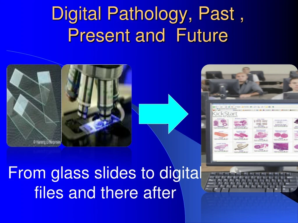
Digital Pathology, Past , Present and Future. From glass slides to digital files and there after. What is Digital Pathology ?. -Is The use of computer technology to convert analog microscopic images into digital images.

Digital Pathology, Past , Present and Future
E N D
Presentation Transcript
Digital Pathology, Past , Present and Future From glass slides to digital files and there after
What is Digital Pathology ? -Is The use of computer technology to convert analog microscopic images into digital images
Other terms are also used such as whole Slide Imaging, abbreviated “WSI”, also known as digital imaging, virtual slides, or virtual microscopy
Introduction Traditionally, education and training in pathology has been delivered using textbooks, glass slides and conventional microscopy. Over the last two decades, the number of web-based pathology resources has expanded dramatically with centralized pathological resources being delivered to many students simultaneously.
Introduction Recently, whole slide imaging technology allows glass slides to be scanned and viewed on a computer screen via dedicated software. This technology is referred to as virtual microscopy and has created enormous opportunities in pathological training and education.
Historical overview of FDA regulation of digital pathology Advances in technology, such as improved image software, scanning throughput, and image storage capabilities have made the wide spread use of digital imaging systems for primary diagnosis a reality. if not now. in the near future.
Historical overview of FDA regulation of digital pathology the use of digital imaging is not new in the clinical or anatomic laboratory---OIVD has cleared and approved several digital imaging devices with limited adjunctive applications.
Historical overview of FDA regulation of digital pathology the use of digital imaging for surgical pathology raises new safety and effectiveness issues that must be addressed. We recognize the many benefits the technology provides, at the same time we need to be sure of it’s limitations to prevent risk to public health.
components in WSI systems -System consisting of hardware; microscope, camera, scanner, computer, and monitor, and software. -Encompasses image acquisition, processing, archiving and retrieval
What does this mean? Conventional optical microscope SLIDE Image Processing software Digital image sensor Mechanical scanner Light source Imaging optics Display READER IMAGE DATA FILES • Microscope one component of the system • Image acquisition, processing and display new technology for this intended use • Diagnostic for neoplastic disease • WSI systems can not be considered Class I exempt
process • Scanning of glass slides by a scanner--- convert information to a digital format • Navigate the digital slide using a software, from any power, on any area of interest, just like a glass slide • Data analysis
Applications Remote access of slides: • Frozen Sections (night calls and remote sites) • Telepathology for 2nd opinion Secondslide.com pathxchange.com • Multi-institution collaborative studies • Retrospective and prospective collections of cases for TMA, case series studies, etc
Real time consultation Bioimagene
Clinical applications • Archival & viewing of data: • Tumor Boards • Quality Assurance (2nd read on QA cases) • Publications (? digital slide access as a part of case reports and case series in the future?) • Scan in key diagnostic slides for outside review cases, to avoid having to borrow back slides for clinical or academic needs. • Access old cases for comparison for frozen sections and current cases • Teaching sets for rare cases.
commercially available WSI instruments • Aperio CSO - Dry scanning and oil immersion • BD KIESTRA™ WCA • Ariol - Clinical IHC and FISH Capture & • -Analysis Digital SlideBox Complete Educational Software
Aperio CSO - Dry scanning and oil immersion Designed specifically for hematopathology and microbiology applications, an Aperio CSO with Oil Immersion uses high numerical aperture oil immersion optics, allowing you to capture the same level of detail when viewing your glass slides under a microscope.
Automated Scoring of Chromogenic Media for the Detection of Methicillin-resistant Staphylococcus aureus (MRSA) Using the WALK-AWAY SPECIMEN PROCESSOR (WASPLab )Image Analysis Software Study about Comparison of automatic imaging to manual detection of MRSA-positive chromogenic agar
Comparison of automatic imaging to manual detection of MRSA-positive chromogenic agar The image taken by the onboard camera is a composite image that uses several light sources and several lighting intensities to simulate manual reading of a plate. Technologists performing manual interpretation used similar images to determine if the plates were positive for MRSA.
In total, 57,690 swabs were enrolled and tested at 4 different locations. The overall prevalence of MRSA was observed to be 2.4% and ranged from 2.1 to 7.3% at the testing sites. Of the 57,690 plates analyzed, 1,367 plates were called positive for MRSA by both automation and manual reading . An additional 5,210 (9.0%) plates that had been manually read as negative were reported as nonnegative by the CDM software. Importantly, the automatic imaging software did not read any manual-positive plates as negative.
Together these data demonstrated an overall sensitivity of 100% (95% CI, 99 to 100%) and a specificity of 90.7% (95% CI, 90 to 91%). Data were similar across all four sites with specificities ranging from 90.0 to 96.0%.
The BD Kiestra™ Work Cell Automation system is a modular solution designed for labs of all sizes that enables the integration of automated specimen processing, plate transportation, incubation and digital imaging systems in a compact footprint. This scalable solution helps to increase productivity and allow your staff to focus more on making clinical decisions and impacting patient care.
Digital IHC From Aperio tutorial,
Ariol - Clinical IHC and Fluorescence in situ hybridization(FISH )Capture & Analysis
Ariol - Clinical IHC and FISH Capture & Analysis The Ariol system, based on the Leica DM6000 B microscope, is the ultimate scalable platform for brightfield, fluorescence and FISH slide scanning and analysis. Ariol combines excellent image capture with quantitative clinical breast panel analysis of Her-2/neu, ER/PR and Her-2/neu FISH. Its unrivalled FISH workflow and image quality on tissue, cells or Tissue slides and tissue microarrays (TMAs), brings FISH review out of the dark room and drastically reduces processing time.
Virtual microscopy and digital pathology in training and education Education and training in histology, anatomical pathology and cytopathology remain essential to both undergraduate and postgraduate courses in pathology and for residency training in pathology. Previously, this would have been delivered using textbooks, glass slides and conventional microscopy, but increasingly web-based resources have been developed to supplement or replace the more traditional methodologies. Web-resources in pathology have expanded dramatically in the last 20 years.
Advantage to Undergraduate From an educational stance, the use of virtual slides also ensures that : - all students see the same slide;that slide is the best and most epresentative in the collection, rather than one of inferior quality, and that poor microscope technique does not interfere with the learning experience.
-It also allows rare slides to be used without fear of breakage. -Displaying the slide on a computer screen means that students can more easily discuss the content with each other, allowing for the use of group-work based approaches to teaching. This pedagogy was more difficult to pursue when students assessed a glass slide on their individual microscopes.
-Virtual microscopy has been shown to improve individual and group learning and enhance the overall learning experience. -Virtual microscopy also has the benefit of delivering courses to students outside the classroom setting. On-line virtual icroscopy courses can be accessed by students nytime, anyplace allowing them to view slides that would have traditionally been restricted to the slide box and the classroom.
Disadvantages. A criticism levelled at virtual microscopy has been that it prevents students from the learning the skills of operating a microscope. This may be true, but in most centres, this is a separate objective to that of analysis and interpretation of tissue sections where the advantages of virtual microscopy clearly outweigh the disadvantages. Indeed, for many groups of students, the ability to operate a microscope successfully is completely irrelevant to their course.
Digital SlideBox Complete Educational Software for Digital Pathology as example The Complete Digital Pathology Education System Digital SlideBox is a world-leading software solution for creating immersive high-impact learning material based on digital pathology slides. Developed in collaboration with leading academic institutes, Digital SlideBox boasts a rich feature set to help bring virtual microscopy into your curriculum.
The software can be accessed from any Internet enabled computer; in class, in the lab and even at home! A flexible structure and in-built custom questionnaires make Digital SlideBox the ideal solution for under graduate, post graduate, continuous professional development (CPD) and external quality assessment (EQA) applications.
Maximize teaching efficiency: -With Digital SlideBox, teaching can be taken out of the microscopy laboratory and onto computers, facilitating increased class sizes and reduced demonstrator overhead costs. -Course content is readily accessible 24/7 from any web-enabled computer, ensuring that students are exposed to course content for longer.
Enrich course content :-Incorporate annotations, gross morphology, radiology images, discussions, web-links, multimedia, and more into your teaching material to provide the most comprehensive learning content to students. -Expose users to rare cases, which they would not traditionally see, using digital pathology, without the risk of breaking valuable glass slides. Standardize content: -Eliminate traditional problems associated with variations in microscope and slide-set quality. -With DSB, every user sees exactly the same content, ensuring that all participants benefit from exposure to the correct information & slides.
Monitor user’s performance -Customized quizzes and integrated multiple choice questionnaires promote independent learning, while instant real-time feedback allows learners to assess their own performance. -In-built heat mapping technology shows the regions and magnification that were observed, giving unique insight into the student’s slide review process.
NEW INTERFACES FOR VIRTUALMICROSCOPY AND EDUCATION The increased use of virtual microscopy for education is driving research to explore new interfaces to manipulate and navigate slides. This closely follows other developments in consumer-based products and their human– computer user interfaces.
A key example of this is multitouch technology which allows individuals to interact with the computer screen, and objects on the screen, by multiple finger touch movements, removing the need for the keyboard or mouse. The authors have developed a multitouch table from Microsoft for the viewing of virtual slides Illustration of the microsoft surface virtual slide ystems developed by Queen’s University Belfast
NEW INTERFACES FOR VIRTUALMICROSCOPY AND EDUCATION Virtual slides can now be viewed using iPad and other tablet devices. PathXL and Leica provide dedicated viewers for virtual microscopy. These will represent important technologies to make histology and histopathology learning more accessible to students
The surface table provides the ability for multiple users to interact simultaneously with a digital slide, again improving group learning in a friendly interactive environment. Touch technology like this is typified by the development of touch screens within handheld smartphones and pad devices. This approach brings enormous benefits to virtual microscopy. A number of companies including Leica and PathXL currently provide iPad/iPhone viewers for virtual microscopy
The Institute for Molecular Medicine Finland has also developed a multitouch large 46- inch screen for digital slide interaction (8). This provides a larger interface to the digital slide. It is likely that there will be an expansion of new multitouch viewing technologies over the next few years.
Practical issues • Cost (machine, software, storage & personal) • Time for scan: 15x15 mm tissue • 20x < 1min; 40 x 6-7 min • Memory: 20X scan: 250 MB; 40X scan: 800 MB • 1TB can hold 2,070 slides (assuming 500 MB/slide), or 4000 slides for 20x (250 MB)
vendors • Aperio • Bioimagene • Olympus (nanozoomer) • Omnyx (UPMC+GE) • 3DHistech • Leica
Issues for evaluation • Scanned image quality • Image management software (interface with laboratory information system (LIS) system if intend for clinical use) • Image analysis software • Technical support • Stability of vendor in the market • Good fit between your need and vendor’s strong points
REFERENCES 1. Harris T, Leaven T, Heidger P, Kreiter C, Duncan J, Dick F. Comparison of a virtual microscope laboratory to a regular microscope laboratory for teaching histology. Anat Rec (New Anat) 001;265:10–4. 2. Heidger PM, Jr, Dee F, Consoer D, Leaven T,Duncan J, Kreiter C. Integrated approach to teaching and testing in histology with real and virtual imaging. Anat Rec (New Anat) 2002; 269:107–12. 3. Krippendorf BB, Lough J. Complete and rapid switch from light microscopy to virtual microscopy for teaching medical histology. Anat Rec (Part B: New Anat) 2005;285B:19–25. 4. Kumar RK, Freeman B, Velan GM, De Permentier PJ. Integrating histology and histopathology teaching in practical classes using virtual slides. Anat Rec (Part B: New Anat)2006;289B:128–33. 5. Scoville SA, Buskirk TD. Traditional and virtual microscopy compared experimentally in a classroom setting. Clin Anat 2007;20:565–70. 5. Lundin J. Youtube Video. http://www.youtube.com/watch?v=woWMgGdzn0M (accessed on 16 January, 2012). 6. Hamilton PW, Bartels PH, Montironi R, Anderson N, Thompson D. Improved diagnostic decision making in pathology. Do inference networks hold the key?. J Pathol 1995;175:1–5.