Chapter 10 The Knee Joint
280 likes | 827 Views
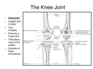
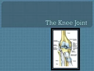
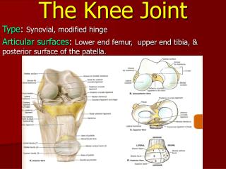
Chapter 10 The Knee Joint. Manual of Structural Kinesiology R.T. Floyd, EdD, ATC, CSCS. The Knee Joint. Knee joint largest joint in body very complex primarily a hinge joint. Bones. Tibia – medial part bears most of weight. Bones. Fibula - lateral

Chapter 10 The Knee Joint
E N D
Presentation Transcript
Chapter 10The Knee Joint Manual of Structural Kinesiology R.T. Floyd, EdD, ATC, CSCS The Knee Joint
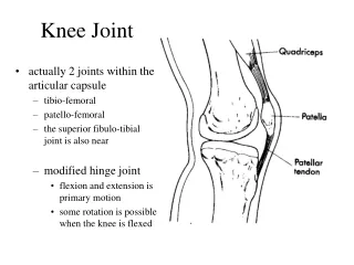
The Knee Joint • Knee joint • largest joint in body • very complex • primarily a hinge joint The Knee Joint
Bones • Tibia – medial part • bears most of weight The Knee Joint
Bones • Fibula - lateral • serves as the attachment for knee joint structures • does not articulate with femur or patella • not part of knee joint The Knee Joint
Bones • Patella • floating bone • imbedded in quadriceps & patellar tendon • serves similar to a pulley in improving angle of pull, resulting in greater mechanical advantage in knee extension The Knee Joint
Bones • Three vasti muscles of quadriceps originate on proximal femur & insert on patellar superior pole • insertion is ultimately on tibialtuberosity via patella tendon • Iliotibial tract of tensor fasciae latae inserts on tibia • Sartorius, gracilis, & semitendinosus insert on upper anteromedialtibial surface The Knee Joint
Bones • Semimembranosus inserts posteromedially on medial tibialcondyle • Biceps femoris inserts primarily on fibula head The Knee Joint
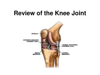
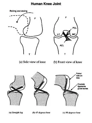
Joints • Articularcartilage surfaces on femur & tibia • Menisci form cushions between bones • attached to tibia • deepen tibialfossa • enhance stability The Knee Joint
Joints • Either or both menisci may be torn causing varying degrees of problems • Tears often occur from significant compression & shear forces during rotation while flexing or extending during quick directional changes in running The Knee Joint
Joints • Anterior & posterior cruciate ligaments • cross within knee between tibia & femur • Important in maintaining anterior & posterior stability, as well as rotational stability • Anterior cruciate ligament (ACL) injuries • one of most common serious injuries to knee • Often caused by noncontact rotational forces with planting & cutting, hyperextension, or by violent quadriceps contraction which pulls tibia forward on femur The Knee Joint
Joints • Extends to 180 degrees (0 degrees of flexion) • Hyperextension of 10 degrees or > not uncommon • Flexion occurs to about 140 degrees The Knee Joint
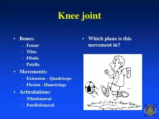
Movements • Flexion • bending or decreasing angle between femur & leg, characterized by heel moving toward buttocks • Extension • straightening or increasing angle between femur & lower leg The Knee Joint
Movements • External rotation • rotary movement of leg laterally away from midline • Internal rotation • rotary movement of lower leg medially toward midline • Neither will occur unless flexed 20-30 degrees or > The Knee Joint
Muscles • Quadriceps muscle group • extends knee • located in anterior compartment of thigh • consists of 4 muscles • rectus femoris • vastus lateralis • vastus intermedius • vastus medialis The Knee Joint
Muscles • Q angle • Central line of pull for entire quadriceps runs from ASIS to the center of patella • Line of pull of patella tendon runs from center of patella to center of tibialtuberosity • Angle formed by the intersection of these two lines at the patella is the Q angle • Generally, females have higher angles due to a wider pelvis The Knee Joint
Muscles • Q angle • Higher Q angles increase the risk for potential knee problems including lateral patellar subluxation or dislocation, chondromalacia, and ACL tears • For people with above normal Q angles, it is important to strengthen the vastusmedialisto counteract the lateral pull of vastuslateralis The Knee Joint
Muscles • Hamstring muscle group • responsible for knee flexion • located in posterior compartment of thigh • consists of 3 muscles • semitendinosus - medial, internal rotator • semimembranosus - medial, internal rotator • biceps femoris - lateral, external rotator The Knee Joint
Rectus Femoris Muscle Flexion of hip Extension of knee Anterior pelvic rotation The Knee Joint
Vastus Lateralis Muscle Extension of knee The Knee Joint
Vastus Intermedius Muscle Extension of knee The Knee Joint
Vastus Medialis Muscle Extension of knee The Knee Joint
Semitendinosus Muscle Flexion of knee Extension of hip Internal rotation of hip Internal rotation of flexed knee Posterior pelvic rotation The Knee Joint
Semimembranosus Muscle Flexion of knee Extension of hip Internal rotation of hip Internal rotation of flexed knee Posterior pelvic rotation The Knee Joint
Biceps Femoris Muscle Flexion of knee Extension of hip External rotation of hip External rotation of flexed knee Posterior pelvic rotation The Knee Joint
Popliteus Muscle Flexion of knee Internal rotation of flexed knee The Knee Joint