
The “ Cellular Internet ”
The “ Cellular Internet ”. Biologists have discovered some universal mechanisms of cellular regulation that involve cell-to-cell communication. External signals are converted into responses within the cell. Methods used by Cells to Communicate. Cell-Cell communication
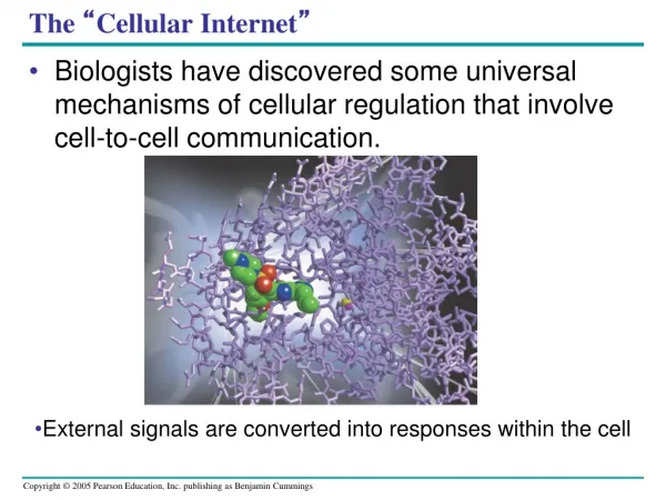

The “ Cellular Internet ”
E N D
Presentation Transcript
The “Cellular Internet” • Biologists have discovered some universal mechanisms of cellular regulation that involve cell-to-cell communication. • External signals are converted into responses within the cell
Methods used by Cells to Communicate • Cell-Cell communication • Cell Signaling using chemical messengers • Local signaling over short distances • Cell-Cell Recognition • Local regulators • Paracrine (growth factors) • Synaptic (neurotransmitters) • Long distance signaling • Hormones
Exchange of mating factors. Each cell type secretes a mating factor that binds to receptors on the other cell type. factor 3 1 2 Receptor a factor Yeast cell, mating type a Yeast cell, mating type Mating. Binding of the factors to receptors induces changes in the cells that lead to their fusion. a New a/ cell. The nucleus of the fused cell includes all the genes from the a and a cells. a/ Figure 11.2 Evolution of Cell Signaling • Yeast cells • Identify their mates by cell signaling
Plasma membranes Plasmodesmata between plant cells Gap junctions between animal cells Figure 11.3 (a) Cell junctions. Both animals and plants have cell junctions that allow molecules to pass readily between adjacent cells without crossing plasma membranes. Cell-Cell Communication • Animal and plant cells • Have cell junctions that directly connect the cytoplasm of adjacent cells
Cell-Cell Communication • Animal cells use gap junctions to send signals • Cells must be in direct contact • Protein channels connecting two adjoining cells Gap junctions between animal cells
Cell-Cell Communication • Plant cells use plasmodesmata to send signals • Cells must be in direct contact • Gaps in the cell wall connecting the two adjoining cells together Plasmodesmata between plant cells
Figure 11.3 (b) Cell-cell recognition. Two cells in an animal may communicate by interaction between molecules protruding from their surfaces. Local Signaling: Cell-Cell Recognition • In local signaling, animal cells may communicate via direct contact • Membrane bound cell surface molecules • Glycoproteins • Glyolipids
Local signaling Target cell Electrical signal along nerve cell triggers release of neurotransmitter Neurotransmitter diffuses acrosssynapse Secretory vesicle Local regulator diffuses through extracellular fluid Target cell is stimulated (b) Synaptic signaling. A nerve cell releases neurotransmitter molecules into a synapse, stimulating the target cell. (a) Paracrine signaling. A secreting cell acts on nearby target cells by discharging molecules of a local regulator (a growth factor, for example) into the extracellular fluid. Local Signaling: Local Regulators • In other cases, animal cells • Communicate using local regulators • Only work over a short distance
Long-distance signaling Blood vessel Endocrine cell Hormone travels in bloodstream to target cells Target cell (c) Hormonal signaling. Specialized endocrine cells secrete hormones into body fluids, often the blood. Hormones may reach virtually all body cells. Figure 11.4 C Long-distance Signaling: Hormones • In long-distance signaling • Both plants and animals use hormones
Long-Distance Signaling • Nervous System in Animals • Electrical signals through neurons • Endocrine System in Animals • Uses hormones to transmit messages over long distances • Plants also use hormones • Some transported through vascular system • Others are released into the air
The Three Stages of Cell Signaling • Earl W. Sutherland (1971) • Discovered how the hormone epinephrine acts on cells • Sutherland suggested that cells receiving signals went through three processes • Reception • Transduction • Response • Called Signal transduction pathways • Convert signals on a cell’s surface into cellular responses • Are similar in microbes and mammals, suggesting an early origin
EXTRACELLULAR FLUID CYTOPLASM Plasma membrane 1 2 3 Reception Transduction Response Receptor Activation of cellular response Relay molecules in a signal transduction pathway Signal molecule Figure 11.5 Overview of cell signaling
Three Stages of Cell Signaling • Signaling molecule binds to the receptor protein CYTOPLASM EXTRACELLULAR FLUID Plasma membrane Reception 1 1 The receptor and signaling molecules fit together (lock and key model, induced fit model, just like enzymes!) Receptor Signaling molecule
Three Stages of Cell Signaling CYTOPLASM EXTRACELLULAR FLUID Plasma membrane Reception Transduction 1 1 2 Receptor 2nd Messenger! Relay molecules in a signal transduction pathway Signaling molecule • The signal is converted into a form that can produce a cellular response
Three Stages of Cell Signaling CYTOPLASM EXTRACELLULAR FLUID Plasma membrane Reception Transduction Response 1 2 3 Receptor Activation of cellular response Relay molecules in a signal transduction pathway Can be catalysis, activation of a gene, triggering apoptosis, almost anything! Signaling molecule • The transduced signal triggers a cellular response
Signal Transduction Animation http://media.pearsoncmg.com/bc/bc_campbell_biology_7/media/interactivemedia/activities/load.html?11&A http://www.wiley.com/legacy/college/boyer/0470003790/animations/signal_transduction/signal_transduction.htm
There are three most common types of membrane receptor proteins. G-protein coupled receptors Receptor tyrosine-kinases Ion channel receptors
What are the different ways that signals can be passed between cells? • Autocrine • Juxtacrine • Paracrine • Endocrine • https://www.dnalc.org/resources/3d/cellsignals.html
1. Reception • A signal molecule, a ligand, binds to a receptor protein in a lock and key fashion, causing the receptor to change shape. Most receptor proteins are in the cell membrane but some are inside the cell. The G-protein is a common membrane receptor.
G-Protein Coupled Receptors are often involved in diseases such as bacterial infections. G-Protein Receptors • https://www.youtube.com/watch?v=V_0EcUr_txk Inactive enzyme Plasma membrane G protein-coupled receptor Activated receptor Signaling molecule Enzyme GDP 2 1 GDP GTP CYTOPLASM G protein (inactive) Activated enzyme i GTP GDP P 4 3 Cellular response
Signal-binding site Signalmolecule Signal molecule Helix in the Membrane Tyr Tyr Tyr Tyr Tyrosines Tyr Tyr Tyr Tyr Tyr Tyr Tyr Tyr Receptor tyrosinekinase proteins(inactive monomers) Dimer CYTOPLASM Activatedrelay proteins Cellularresponse 1 Tyr Tyr Tyr Tyr Tyr Tyr P P Tyr P Tyr Tyr Tyr Tyr P Tyr Tyr Tyr P P P Tyr Tyr Tyr Tyr Tyr P Tyr Tyr Tyr Cellularresponse 2 P P P Tyr Tyr P 6 ATP 6 ADP Activated tyrosine- kinase regions (unphosphorylated dimer) Fully activated receptor tyrosine-kinase (phosphorylated dimer) Inactiverelay proteins Figure 11.7 Tyrosine Kinase model video • Receptor tyrosine kinases normal and wrong cancer and tk
Ion Channel Receptors Gate closed 1 Ions Signaling molecule (ligand) • Very important in the nervous system • Signal triggers the opening of an ion channel • depolarization • Triggered by neurotransmitters Ligand-gated ion channel receptor Plasma membrane 2 Gate open Cellular response 3 Gate closed
2. Transduction • Transduction: Cascades of molecular interactions relay signals from receptors to target molecules in the cell • Multistep pathways • Can amplify a signal (Amplifies the signal by activating multiple copies of the next component in the pathway) • Provide more opportunities for coordination and regulation • At each step in a pathway, the signal is transduced into a different form, commonly a conformational change in a protein.
Signaling molecule Transduction: A Phosphorylation Cascade Receptor Activated relay molecule Inactive protein kinase 1 Fig. 11-9 Active protein kinase 1 Inactive protein kinase 2 ATP Phosphorylation cascade ADP P Active protein kinase 2 PP P i Inactive protein kinase 3 ATP ADP P Active protein kinase 3 PP P i Inactive protein ATP P ADP Active protein Cellular response PP P i
Protein Phosphorylation and Dephosphorylation • Many signal pathways • Include phosphorylation cascades • In this process, a series of protein kinases add a phosphate to the next one in line, activating it • Phosphatase enzymes then remove the phosphates
Signal molecule A relay molecule activates protein kinase 1. Receptor Activated relay molecule 4 1 3 5 2 Inactive protein kinase 1 Active protein kinase 1 transfers a phosphate from ATP to an inactive molecule of protein kinase 2, thus activating this second kinase. Active protein kinase 1 Active protein kinase 2 then catalyzes the phos- phorylation (and activation) of protein kinase 3. Inactive protein kinase 2 ATP Phosphorylation cascade P Active protein kinase 2 ADP PP P i Enzymes called protein phosphatases (PP) catalyze the removal of the phosphate groups from the proteins, making them inactive and available for reuse. Inactive protein kinase 3 Finally, active protein kinase 3 phosphorylates a protein (pink) that brings about the cell’s response to the signal. ATP P ADP Active protein kinase 3 PP P i Inactive protein ATP P ADP Active protein Cellular response PP P i • A phosphorylation cascade Figure 11.8
The transduction stage of signaling is often a multistep process that amplifies the signal. About 1% of our genes are thought to code for kinases.
Small Molecules and Ions as Second Messengers • Secondary messengers • Are small, nonprotein, water-soluble molecules or ions that act as secondary messengers.
First messenger (signal molecule such as epinephrine) Adenylyl cyclase G protein GTP G-protein-linked receptor ATP cAMP Protein kinase A Cellular responses Cyclic AMP http://media.pearsoncmg.com/bc/bc_campbell_biology_7/media/interactivemedia/activities/load.html?11&C • Many G-proteins trigger the formation of cAMP, which then acts as a second messenger in cellular pathways. Figure 11.10
NH2 NH2 NH2 N N N N N N N N N N N O O O N O Adenylyl cyclase Phoshodiesterase CH2 O HO Ch2 P –O O P O P P O CH2 O O O O O O O O O P Pyrophosphate H2O O O P P i OH OH OH OH OH ATP Cyclic AMP AMP Cyclic AMP • Cyclic AMP (cAMP) • Is made from ATP
First messenger Adenylyl cyclase G protein Fig. 11-11 GTP G protein-coupled receptor ATP Second messenger cAMP Transduction in a G-protein pathway Protein kinase A Cellular responses
EXTRACELLULAR FLUID Plasma membrane Ca2+pump ATP Mitochondrion Nucleus CYTOSOL Ca2+pump Endoplasmic reticulum (ER) ATP Ca2+pump Key High [Ca2+] Low [Ca2+] Calcium ions and Inositol Triphosphate (IP3) • Calcium, when released into the cytosol of a cell acts as a second messenger in many different pathways Calcium is an important second messenger because cells are able to regulate its concentration in the cytosol Other second messengers such as inositol triphosphate and diacylglycerol can trigger an increase in calcium in the cytosol
6 3 2 1 4 5 A signal molecule binds to a receptor, leading to activation of phospholipase C. DAG functions as a second messenger in other pathways. Phospholipase C cleaves a plasma membrane phospholipid called PIP2 into DAG and IP3. EXTRA- CELLULAR FLUID Signal molecule (first messenger) G protein DAG GTP PIP2 G-protein-linked receptor Phospholipase C IP3 (second messenger) IP3-gated calcium channel Endoplasmic reticulum (ER) Various proteins activated Cellularresponse Ca2+ Ca2+ (second messenger) The calcium ions activate the next protein in one or more signaling pathways. IP3 quickly diffuses through the cytosol and binds to an IP3– gated calcium channel in the ER membrane, causing it to open. Calcium ions flow out of the ER (down their con- centration gradient), raising the Ca2+ level in the cytosol. http://www.ncbi.nlm.nih.gov/books/NBK10883/figure/A247/?report=objectonly Figure 11.12
Growth factor 3. Response Receptor Reception • Many possible outcomes • This example shows a transcription response Phosphorylation cascade Transduction CYTOPLASM Inactive transcription factor Active transcription factor Response P DNA Gene NUCLEUS mRNA
Signaling molecule • Specificity of the signal • The same signal molecule can trigger different responses • Many responses can come from one signal! Receptor Relay molecules Response 1 Response 2 Response 3 Cell A. Pathway leads to a single response. Cell B. Pathway branches, leading to two responses.
http://www.hhmi.org/biointeractive/signal-molecules-trigger-transcription-factorshttp://www.hhmi.org/biointeractive/signal-molecules-trigger-transcription-factors • The signal can also trigger an activator or inhibitor • The signal can also trigger multiple receptors and different responses Activation or inhibition Response 4 Response 5 Cell C. Cross-talk occurs between two pathways. Cell D. Different receptor leads to a different response.
Response- cell signaling leads to regulation of transcription (turn genes on or off) or cytoplasmic activities.
Long-distance SignalingIntracellular signaling includes hormones that are hydrophobic and can cross the cell membrane. Once inside the cell, the hormone attaches to a protein that takes it into the nucleus where transcription can be stimulated. Testosterone acts as a transcription factor.
Hormone (testosterone) EXTRACELLULAR FLUID 1 The steroid hormone testosterone passes through the plasma membrane. Plasma membrane Receptor protein 2 Testosterone binds to a receptor protein in the cytoplasm, activating it. Hormone- receptor complex 3 The hormone- receptor complex enters the nucleus and binds to specific genes. DNA mRNA 4 The bound protein stimulates the transcription of the gene into mRNA. NUCLEUS New protein 5 The mRNA is translated into a specific protein. CYTOPLASM Figure 11.6 • Steroid hormones • Bind to intracellular receptors
Signalmolecule Plasmamembrane Receptor Threedifferentproteinkinases Scaffoldingprotein Figure 11.16 Signaling Efficiency: Scaffolding Proteins and Signaling Complexes • Scaffolding proteins • Can increase the signal transduction efficiency
Termination of the Signal • Signal response is terminated quickly • By the reversal of ligand binding
Any Questions?? Can You Hear Me Now?
Two systems control all physiological processes 1. Nervous System – neurosecretory glands in endocrine tissues secrete hormones. 2. Endocrine System
Major Vertebrate Endocrine Glands Their Hormones (Hypothalamus–Parathyroid glands)
Neurosecretory cells in endocrine organs and tissues secrete hormones. These hormones are excreted into the circulatory system.
Stress and the Adrenal Gland http://highered.mcgraw-hill.com/olcweb/cgi/pluginpop.cgi?it=swf::535::535::/sites/dl/free/0072437316/120109/bio48.swf::Action%20of%20Epinephrine%20on%20a%20Liver%20Cell
http://bcs.whfreeman.com/thelifewire/content/chp42/4202003.htmlhttp://bcs.whfreeman.com/thelifewire/content/chp42/4202003.html


