Download
1 / 32
330 likes | 475 Views
Lecture 4. Mass spectrometric methods. Assistant of the pharmaceutical chemistry department Burmas Nataliya Ivanivna e-mail: Natashenka-Burmas@rambler.ru. Plan. The concept of mass-spectrometric analysis. The mass spectrometer Quantitative and qualitative analysis

E N D
Lecture 4 Mass spectrometric methods Assistant of the pharmaceutical chemistry department Burmas Nataliya Ivanivna e-mail: Natashenka-Burmas@rambler.ru
Plan • The concept of mass-spectrometric analysis. • The mass spectrometer • Quantitative and qualitative analysis • Applications of mass-spectrometry
The concept of mass-spectrometry analysis Mass spectrometry (MS) is an analytical technique for the determination of the elemental composition of a sample or molecule. It is also used for elucidating the chemical structures of molecules, such as peptides and other chemical compounds.
In a typical MS procedure: • a sample is loaded onto the MS instrument, and undergoes vaporization. • the components of the sample are ionized by one of a variety of methods (e.g., by impacting them with an electron beam), which results in the formation of charged particles (ions) • the positive ions are then accelerated by an electric field • computation of the mass-to-charge ratio (m/z) of the particles based on the details of motion of the ions as they transit through electromagnetic fields, and • detection of the ions, which in step 4 were sorted according to m/z.
Analysers characteristics The mass resolving power - is the measure of the ability to distinguish two peaks of slightly different m/z. The mass accuracy - is the ratio of the m/z measurement error to the true m/z. Usually measured in ppm or milli mass units. The mass range - is the range of m/z amenable to analysis by a given analyzer. The linear dynamic range - is the range over which ion signal is linear with analyte concentration.
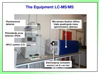
2. The mass spectrometer In order to measure the characteristics of individual molecules, a mass spectrometer converts them to ions so that they can be moved about and manipulated by external electric and magnetic fields.
The word spectrograph has been used since 1884 as an "International Scientific Vocabulary". The linguistic roots are a combination and removal of bound morphemes and free morphemes which relate to the terms spectrum and photographic plate. Early spectrometry devices that measured the mass-to-charge ratio of ions were called mass spectrographs which consisted of instruments that recorded a spectrum of mass values on a photographic plate. A mass spectroscope is similar to a mass spectrograph except that the beam of ions is directed onto a phosphor screen.
The three essential functions of a mass spectrometer, and the associated components, are: • A small sample of compound is ionized, usually to cations by loss of an electron. (the ion source) • The ions are sorted and separated according to their mass and charge. (the mass analyzer). • The separated ions are then detected and tallied, and the results are displayed on a chart. (the detector)
The analyser and detector of the mass spectrometer, and often the ionisation source too, are maintained under high vacuum to give the ions a reasonable chance of travelling from one end of the instrument to the other without any hindrance from air molecules. The entire operation of the mass spectrometer, and often the sample introduction process also, is under complete data system control on modern mass spectrometers.
The technique has both qualitative and quantitative uses. These include identifying unknown compounds, determining the isotopic composition of elements in a molecule, and determining the structure of a compound by observing its fragmentation. Other uses include quantifying the amount of a compound in a sample or studying the fundamentals of gas phase ion chemistry (the chemistry of ions and neutrals in a vacuum).
Because ions are very reactive and short-lived, their formation and manipulation must be conducted in a vacuum. Atmospheric pressure is around 760 torr (mm of mercury). The pressure under which ions may be handled is roughly 10-5 to 10-8 torr (less than a billionth of an atmosphere). Each of the three tasks listed above may be accomplished in different ways. In one common procedure, ionization is effected by a high energy beam of electrons, and ion separation is achieved by accelerating and focusing the ions in a beam, which is then bent by an external magnetic field. The ions are then detected electronically and the resulting information is stored and analyzed in a computer.
When a high energy electron collides with a molecule it often ionizes it by knocking away one of the molecular electrons (either bonding or non-bonding). This leaves behind a molecular ion (colored red in the following diagram). Residual energy from the collision may cause the molecular ion to fragment into neutral pieces (colored green) and smaller fragment ions (colored pink and orange). The molecular ion is a radical cation, but the fragment ions may either be radical cations (pink) or carbocations (orange), depending on the nature of the neutral fragment.
Many mass spectrometers work in either negative ion mode or positive ion mode. It is very important to know whether the observed ions are negatively or positively charged. This is often important in determining the neutral mass but it also indicates something about the nature of molecules.
bromine Bromine: 50.50% 79Br and 49.50% 81Br
Isotopes Since a mass spectrometer separates and detects ions of slightly different masses, it easily distinguishes different isotopes of a given element. This is manifested most dramatically for compounds containing bromine and chlorine, as illustrated by the following examples. Since molecules of bromine have only two atoms, the spectrum on the left will come as a surprise if a single atomic mass of 80 amu is assumed for Br. The five peaks in this spectrum demonstrate clearly that natural bromine consists of a nearly 50:50 mixture of isotopes having atomic masses of 79 and 81 amu respectively. Thus, the bromine molecule may be composed of two 79Br atoms (mass 158 amu), two 81Br atoms (mass 162 amu) or the more probable combination of 79Br-81Br (mass 160 amu). Fragmentation of Br2 to a bromine cation then gives rise to equal sized ion peaks at 79 and 81 amu.
vinyl chloride Chlorine: 75.77% 35Cl and 24.23% 37Cl
3. Quantitative and qualitative analysis Mass analyzers separate the ions according to their mass-to-charge ratio. The following two laws govern the dynamics of charged particles in electric and magnetic fields in vacuum: (Lorentz force law) where F is the force (in newtons) E is the electric field (in volts per meter) B is the magnetic field (in teslas) q is the electric charge of the particle (in coulombs) v is the instantaneous velocity of the particle (in meters per second) × is the vector cross product
Newton's second law of motion in non-relativistic case, i.e. valid only at ion velocity much lower than the speed of light. Here F is the force applied to the ion, m is the mass of the ion, a is the acceleration
Many mass spectrometers work in either negative ion mode or positive ion mode. It is very important to know whether the observed ions are negatively or positively charged. This is often important in determining the neutral mass but it also indicates something about the nature of molecules. Different types of ion source result in different arrays of fragments produced from the original molecules.
By understanding the origin of a sample, certain expectations can be assumed as to the component molecules of the sample and their fragmentations. A sample from a synthesis/manufacturing process will probably contain impurities chemically related to the target component. A relatively crudely prepared biological sample will probably contain a certain amount of salt, which may form adducts with the analyte molecules in certain analyses.
4. Application of mass-spectrometry Mass spectrometry is also used to determine the isotopic composition of elements within a sample. Differences in mass among isotopes of an element are very small, and the less abundant isotopes of an element are typically very rare, so a very sensitive instrument is required. These instruments, sometimes referred to as isotope ratio mass spectrometers (IR-MS), usually use a single magnet to bend a beam of ionized particles towards a series of Faraday cups which convert particle impacts to electric current.
A fast on-line analysis of deuterium content of water can be done using Flowing afterglow mass spectrometry, FA-MS. Probably the most sensitive and accurate mass spectrometer for this purpose is the accelerator mass spectrometer (AMS). Isotope ratios are important markers of a variety of processes. Some isotope ratios are used to determine the age of materials for example as in carbon dating. Labeling with stable isotopes is also used for protein quantification.