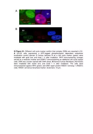
Subcellular Localization of GFP-OsCAF1 Fusion Proteins in Onion Epidermal Cells
This supplementary figure shows the subcellular localization of different OsCAF1 proteins fused to GFP in onion epidermal cells. The green fluorescence signals emitted from GFP were detected using a fluorescence microscope. Nuclei are marked by white arrows. Scale bar = 50 μm.
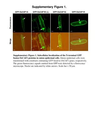

Subcellular Localization of GFP-OsCAF1 Fusion Proteins in Onion Epidermal Cells
E N D
Presentation Transcript
Supplementary Figure 1. GFP-OsCAF1A GFP-OsCAF1B (L) GFP-OsCAF1G GFP-OsCAF1H Fluorescence Merge Supplementary Figure 1. Subcellular localization of the N-terminal GFP fusion OsCAF1 proteins in onion epidermal cells. Onion epidermal cells were transformed with constructs containing GFP fused to OsCAF1genes, respectively. The green fluorescence signals emitted from GFP were detected by a florescence microscope. Nuclei are indicated by white arrows. Scale bar = 50 μm.