Download
1 / 80
800 likes | 957 Views
Acute Coronary Syndromes. Dr. S. McPherson Dr. G. Curry July 11, 2002. Chest Pain – the initial presentation. 2 questions to be answered: Are the symptoms a manifestation of an ACS? If so, what is likelihood of an adverse outcome?. Chest pain – the initial presentation.

E N D
Acute Coronary Syndromes Dr. S. McPherson Dr. G. Curry July 11, 2002
Chest Pain – the initial presentation 2 questions to be answered: Are the symptoms a manifestation of an ACS? If so, what is likelihood of an adverse outcome?
Chest pain – the initial presentation • Early Risk stratification: • Categorize patients with chest pain into high, intermediate, and low risk of ACS • Use of history (anginal symptoms), physical exam, ECG, biochemical markers
Historical features Classic characteristics: • Deep chest or arm pain associated with exertion or emotional stress • Resolves promptly with rest or NTG Not characteristic: • Pleuritic • Primarily mid to lower abdominal pain • Pain that is localized with 1 finger • Reproducible pain with palpation or movement • Constant pain X hours • Very brief pain lasting few seconds or less • Pain radiates to lower extremities
Incidence of ACS with noncharacteristic symptoms Arch Int Med 1985;145:65-9 • Sharp or stabbing pain 22% • Pleuritic 13% • Reproducible to palpation 7% Med Care 1991; 29:610-27, J Critic epi 1992;85:1254-64 • Traditional risk factors only weakly predictive of ACS • But presence of risk factors relate to poorer outcomes
Estimation of the risk of adverse outcomes JAMA 1996;276:1568-74 • Looked at 30 day mortality or MI in patients with high, intermediate and low risk UA (AMI and noncardiac CP excluded) • Low risk 0% • Intermediate risk 1.2% • High risk 1.7%
Management recommendations • Low risk: expeditious work up as an outpatient (ie within 72 hrs of d/c from ED) • High and intermediate risk admit for further work up
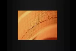
The ECG • Progression of changes • Giant R wave • Hyperacute T waves (release of intracellular K+) • ST elevation (current of injury) • Q wave (shift of axis of depolarization) • Inverted T waves (abnormal repolarization
Interpreting the ECG • ECG • Inferior II, III, aVf • Lateral I, aVL, V5-6 • Anterior V1-4 • Posterior ST depression ant V1-3 with large R V1
Interpreting the ECG • ECG in association with old LBBB (Sgarbossa) any concordant change >1mm any discordant change > 5mm *same applies for VPB rhythm • 15 lead ECG prognostic value sensitivity
Interpreting the ECG • ST elevation of > 0.1 mV confirmed >90% of the time by biochemical markers to be AMI • Up to 15% of AMI have a normal or nonspecific ECG • ST depression of > 0.05 mV or T wave inversion of > 0.2 mV may represent cardiac ischemia
Biochemical Cardiac Markers • Lab • CK, CKMB • At 24Hrs sensitivity reaches >95% • Specificity problem (high false positive rate) • Doesn’t predict USA • Troponins (TnT, TnI) • T1/2 : 3-8 hrs, but detected for up to 10 days
Biochemical markers • Enzyme comparison Markertime to peak time to normmyoglobin 1-3 6-10 24-36 CK 3-8 10-36 72-96 CK-MB 3-8 9-30 48-72 CK-MB iso 1-3 4-6 18-24 cTnT 2-6 10-24 10-15 days cTnI 2-6 12-24 7-10 days
Troponin the main points • Troponin should NOT determine disposition • General Numbers to remember • 6 -----------------> 60% sensitive • 8 -----------------> 80% sensitive • 10 -----------------> 90% sensitive • Patients with TnT + and CKMB – have worse prognosis (increased death and CV events at 30 days)
False + troponin • Troponins: increase levels? • Ischemia/ infarction • Cardiomyopathy • Electrical injury • Pericarditis • Myocarditis • Cardiac contusion • Hypertensive emergencies • End- stage renal failure • Pulmonary embolus
Case 1 & 2 • Insert a low risk ACS case and a high or intermediate case and have group decide what to do in light of the previous slides
Unstable angina/NSTEMI Canadian Cardiology Classification of Angina • Class I. Ordinary physical activity (e.g., walking or climbing stairs) does not cause angina. • Class II. Slight limitation of normal activity; angina occurs with walking, climbing stairs, or emotional stress. • Class III. Severe limitation of ordinary physical activity; angina occurs on walking one or two blocks on a level surface or climbing one flight of stairs in normal conditions. • Class IV. Inability to carry on any physical activity without discomfort; anginal symptoms may be present at rest.
USA/NSTEMI • Definition of Unstable Angina: Rest angina. Angina occurring at rest, lasting longer than 20 minutes, and occurring within 1 week of presentation New-onset angina. Angina of at least class III severity with onset within the last 2 months Increasing angina. Previously diagnosed angina that is more frequent, longer in duration, or increased by one class within the last 2 months of at least class III severity • Definition of NSTEMI: Evidence of myocardial necrosis with rise in biochemical markers
Case # 3 • Insert case of classic USA here
Anti-ischemic therapy: Bedrest Oxygen NTG Morphine B-blocker CCB ACE Anti-platelet/Anti-thrombotic therapy: ASA Clopidogrel GPIIbIIIA Heparin LMWH Management of UA/NSTEMI
Anti-ischemic therapy • Oxygen • No evidence to support administration of O2 to all patients with ACS • Recommended for patients with questionable respiratory status and hypoxia
Anti-ischemic therapy Nitrates: • 0.4mg sl or spray q5min • Initiate NTG infusion if above does not resolve pain (contraindicated if hypotension or pt has used Viagra within 24hr); titrate q3min to effect or until SBP < 110mmHg
Nitrates – the evidence • Meta-analysis of the small trials (pre-lytic) showed a 35% reduction in mortality • ISIS-4 and GISSI-3 showed no mortality benefit but frequent pre-hospital and hospital use of NTG in the control group • BOTTOM LINE: we all use it, it makes patients feel better, it doesn’t seem to harm most patients
Anti-ischemic therapy Morphine: • 1-5 mg iv recommended for patients with ongoing pain post NTG x3 • May give with iv NTG but watch BP • No RTC to validate its benefit or clarify its optimal schedule • Adverse effects: hypotension, N&V (20%), resp depression
Anti-ischemic therapy B-Adrenergic blockers: • Metoprolol 5mg iv q5min, if tolerate 15mg start 25mg po 15min after last iv dose • Contrindications: • 1st, 2nd, 3rd degree heart block, LV dysfunction, CHF, asthma, sinus brady, hypotension, cautiously in patients with COPD
B-blockers the evidence • Overview of all RPCT (small) showed 13% reduction is progression to AMI, not powered to detect difference if mortality JAMA 1988 • Use in USA extrapolated from mortality reduction proven in AMI, stable angina, heart failure
Anti-ischemic therapy Calcium channel blockers: • To control ongoing or recurrent pain when maxed therapy of B-blocker/NTG, NTG/B-blocker not tolerated or variant angina • Do not use nifedipine • Do not use verapamil or diltiazem if evidence of acute CHF • Evidence supports role for symptom control with use as a rate control if B-blocker not tolerated
UA/NSTEMI – Antiplatelet therapy • ASA • Clopidogrel • GPIIbIIa inhibitors
Anti-platelet therapy ASA: • 160-325mg to chew ASAP • All trials have shown mortality benefit that extends to 2 years • Contraindications: • Allergy • Active bleeding (GI, retinal) • Hemophilia
Anti-platelet therapy Clopidogrel • CAPRIE (1996), RCT ASA vs Plavix (N=19,185) • 3 year ischemic stroke, MI or death ARR = 0.5% (NNT = 200) • Plavix equal to ASA but increased side effects (diarrhea, rash, GI bleed)
Anti-platelet therapy Clopidogrel: • CURE trial (2001) RCT Plavix + ASA vs ASA • UA/NSTEMI within 24 hr • Death, MI, or stroke 3 and 12 month ARR = 2.2% (NNT = 45) • Excess in major bleed of 1% (NNH = 100) • Risk of bleed with CABG increased in 1st 5 days • Recommended in UA/NSTEMI when noninvasive course is anticipated • BOTTOM LINE: appears to work but not for us to start in the ED
Anti-platelet therapy GPIIbIIIa Receptor Antagonists: • Inhibits the cross-linking of platelets by fibrinogen
GPIIbIIIa RA • EPIC (abcixamab) NEJM 1994 • PRCT, N=2,099 pts • High risk pts going for PCI (AMI, USA) • Reopro(bolus, bolus +infusion) vs placebo • Placebo vs bolus similar • F/U 30 days • Bolus/infusion: lower rate triple end-point (Death, MI or urgent revascularization) 8.3% vs 12.8% (ARR 4.5%, RRR 35%) • 2 fold increase in major bleeds: 14% vs 6.6 (ARI 7.4%, RRI 112%!!!
GPIIbIIIa RA • CAPTURE (abcixamab) Lancet 1997 • PRCT, N=1050 pts (stopped early) • Pts with refractory USA, after cath and before plasty • Reopro vs placebo , 18-24hrs before plasty, and 1 hr after • F/U 30 days then 6 months • Composite endpoint at 30 days(Death, MI, urgent intervention ): 2.6% vs 5.5% (ARR 2.9%, RRR 53%) • At 6 months: no difference! (decreased early events only) • Major bleeding: 3.8% vs 1.9% (ARI 1.9%, RRI 50%)
GPIIbIIIa RA • CAPTURE (substudy) • Predictive value of TnT • Increased TnT(>0.1 ng/ml): 30.9% • F/U 6 months: Death or MI • TnT +ve • Reopro 9.5% vs placebo 23.9% (ARR 14.4%, RRR 60% • TnT –ve • Reopro 7.5% vs 9.4% (not significant) • Conclusion: TnT +ve identifies a high risk population that seems to benefit from GIIb-IIIa’s
GPIIbIIIa RA • PRISM-PLUS (tirofiban) NEJM 1998 • PRCT, N=1,570 pts • Non-ST segment elevation ACS and likely to go to catheterization • Heparin vs tirofiban vs combination • Tirofiban alone arm stopped (increased mortality at 7 days) • F/U 7 and 30 days and 6 months • Composite endpoint (Death, MI or recurrent ischemia): 7 days: 12.9 vs 17.9% (p=0.004), 30 days: not significant, 6 months: 27.7 vs 32.2% (p=0.02)
GPIIbIIIa RA • PURSUIT (eptifibatide) • PRCT, N=10,948 pts • Eligible if ECG changes : transient ST elevation, ST depression, T wave inversion, or positive CKMB • Eptifibitide vs placebo • F/U 30 days and 6 months • Double endpoint (Death or MI): 30 days: 14.2 vs 15.7% (ARR 1.5%, RRR 10%), 6 months: 12.1 vs 13.6% (ARR 1.5%, RRR 11%) • Medical Management: US benefit, Europe and Latin America no benefit • Didn’t work in females!!!!!
GPIIbIIIa RA • GUSTO IV • PRCT, N=7800 pts • ECG changes, +ve troponin level • Early revascularization strongly discouraged • Abcixamab vs placebo (24-48 hr infusion) • No difference in MI or Death
GPIIbIIIa RA BOTTOM LINE: • They are definitely indicated as an adjunct to PCI • Role in UA/NSTEMI is questionable, there appears to be a small reduction in death or MI in high risk groups (ongoing or recurrent pain, dynamic ST changes) • Not enough evidence for us to start in the ED
Anti-thrombotic therapy Unfractionated Heparin: • 5000 Unit bolus then 1000 unit/hr • Evidence is scanty: • Theroux et al (1993) RCT ASA+hep vs ASA • Decreased rates of MI (fatal and nonfatal) 3% (NNT = 33%) • RISC (1990) ASA vs UFH vs UFH +ASA • ASA + UFH had lowest events in 1st 5 days
Anti-thrombotic therapy LMWH: • ESSENCE (1997) RCT enoxaparin vs UFH • No difference in mortality • Composite endpoint of death, MI or recurrent angina, ARR = 3.2% (NNT=33) • TIMI 11B (1999) RCT enoxaparin vs UFH • Composite endpoint death, MI, urgent need for PTCA, ARR = 2.1% (NNT = 50) • 2 trials (1 dalteparin, 1 nadroparin) had neutral or unfavorable trends
Case #4 • Insert case for AMI here
Diagnosis of AMI • WHO def’n (2 of 3): • Ischemic type chest discomfort • Rise & fall in serum cardiac markers • Changes on ECG • ST elevation 91% specific, 46% sensitive for MI
Management of AMI • Routine measures: • Supplemental O2 • Iv access • ECG interpreted within 10 minutes of arrival to ED