
Structural Study of Thermolysin-Cbz-GlyP-Leu-Leu Complex: Inhibitor Binding Mechanism
Explore the 3D structure of the complex formed by thermolysin and Cbz-GlyP-Leu-Leu, analyzing how the inhibitor binds and impacts the protein's active sites. Learn about the strategic placement of Leucine residues and inhibitor conformation.

Structural Study of Thermolysin-Cbz-GlyP-Leu-Leu Complex: Inhibitor Binding Mechanism
E N D
Presentation Transcript
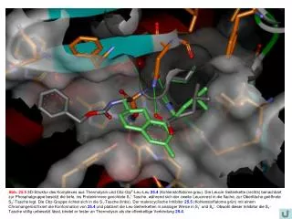
Abb. 25.5 3D-Struktur des Komplexes aus Thermolysin und Cbz-GlyP-Leu-Leu 25.4 (Kohlenstoffatome grau). Die Leucin-Seitenkette (rechts) benachbart zur Phosphatgruppe besetzt die tiefe, ins Proteininnere gerichtete S1’-Tasche, während sich der zweite Leucinrest in die flache, zur Oberfläche geöffnete S2’-Tasche legt. Die Cbz-Gruppe richtet sich in die S1-Tasche (links). Der makrocyclische Inhibitor 25.5 (Kohlenstoffatome grün) mit einem Chromangerüst fixiert die Konformation von 25.4 und platziert die Leu-Seitenketten in analoger Weise in S1’ und S2’. Obwohl dieser Inhibitor die S1-Tasche völlig unbesetzt lässt, bindet er fester an Thermolysin als die offenkettige Verbindung 25.4.