Download

1 / 29
290 likes | 324 Views
Delve into the realm of small units in science, from molecules with molecular weights to cells varying in size, using the metric system. Discover what makes up the smallest components of life, from ribosomes to organic acids. Learn about prokaryotic cells' structures and sizes, comparing Gram-negative and Gram-positive bacteria.

E N D
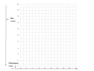
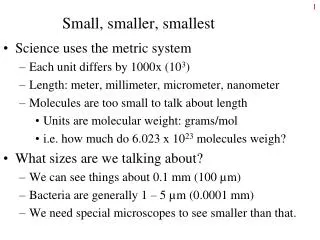
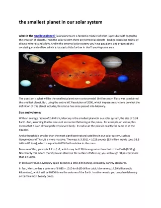
Small, smaller, smallest • Science uses the metric system • Each unit differs by 1000x (103) • Length: meter, millimeter, micrometer, nanometer • Molecules are too small to talk about length • Units are molecular weight: grams/mol • i.e. how much do 6.023 x 10²³ molecules weigh? • What sizes are we talking about? • We can see things about 0.1 mm (100 µm) • Bacteria are generally 1 – 5 µm (0.0001 mm) • We need special microscopes to see smaller than that.
Something can’t be smaller than the parts it is made of! Sand is used to make bricks, and bricks are assembled to make a house. A house can’t be smaller than a brick; a brick can’t be smaller than grains of sand. Likewise, small molecules are combined to make polymers and polymers are used to make cells. cic.nist.gov/lipman/sciviz/scan/jun24_ptC1a.jpg www.littleitalymd.com/images/BrickLayer.gif www.kevscartoons.com/images/illustration/chil...
In the world of small, what’s big? • Cells of eukaryotic organisms are big • Nerve cells can be quite long • White blood cells are about 10 µm in diameter • An amoeba may be around 20 µm • Prokaryotes and cell organelles are smaller • E. coli is about 1 µm long • A mitochondrion is about the same size • Particles are smaller • Viruses range from 20 to 200 nm (0.02 – 0.2 µm) • Ribosomes, found inside cells, are about 20 nm
In the world of small, what’s smaller? • Ribosomes, viruses, cell walls are made of polymers • Ribosomes and viruses are combinations of proteins and nucleic acids • Cell walls are made of large molecules like peptidoglycan and lipopolysaccharide • Polymers are larger than the monomers they are made of • Proteins range from 10,000 to 500,000 MW • Bacterial DNA is over 1 mm long! (but very skinny) • Polysaccharides can be > 100,000 MW (grams/mol)
In the world of small, what’s smallest? • These are all small molecules ranging from 18 g/mol to 1,000 g/mol • Water, oxygen gas, nitrogen gas • Sugars (glucose, sucrose, etc.) • Amino acids • Nucleotides • Fatty acids, cholesterol, (even phospholipids aren’t big) • Organic acids found in metabolism • Vitamins • Antibiotics and most other drugs
Life and Cells • What is Life? • Can grow, i.e. increase in size. • Can reproduce. • Responsive to environment. • Metabolism: can acquire and utilize energy. • Schwann and Schleiden: cells basic unit of life • Prokaryotes and eukaryotes from microscopy. • Our focus: prokaryotic cells. • Eubacteria and Archaebacteria
Bacterial Appearance • Size • 0.2 µm – 0.1 mm • Most 0.5 – 5.0 µm • Shape • Coccus (cocci); rod (bacillus, bacilli); spiral shapes (spirochetes; spirillum, spirilla); filamentous; various odd shapes. • Arrangement • Clusters, tetrads, sarcina, pairs, chains http://www.cellsalive.com/howbig.htm http://www.ionizers.org/Sizes-of-Bacteria.html http://smccd.net/accounts/case/biol230/ex3/bact.jpeg
Overview of prokaryotic cell.
From Membrane Out:lecture order • Examination of layers of bacterial cell • Starting at cell membrane, working to outside • A look at how cells move • Examination of inside of bacterial cell • A look at how things get into cells • Brief review of eukaryotic cell structure.
Structure of phospholipids http://biyoloji_genetik.sitemynet.com/genel_biyoloji/genel_biyoloji_logos/phospholipids.gif
How phospholipids work Polar head groups associate with water but hydrophobic tails associate with each other to avoid water. When placed in water, phospholipids associate spontaneously side by side and tail to tail to form membranes. http://users.rcn.com/jkimball.ma.ultranet/BiologyPages/L/LipidBilayer.gif
Cell Membranes • 50/50 lipids and proteins • Fluid mosaic model • Effective barrier to large and hydrophilic molecules • O2, CO2, H2O, lipid substances can pass through • Salts, sugars, amino acids, polymers, cannot. • Proteins can be on inner, outer surfaces (peripheral) or transmembrane (integral) • Involved primarily with transport • Degradation and biosynthesis • Site of ATP synthesis
Membrane structure http://www.slic2.wsu.edu:82/hurlbert/micro101/images/cytomemb.gif
Outside the cell membrane:the Cell Wall Animal cells do not have a cell wall outside the cell membrane. Plant cells and fungal cells do. So do most prokaryotic cells, providing structural support and influencing the shape of the cell.
Division of the Eubacteria:Gram Negative and Gram Positive • Gram stain invented by Hans Christian Gram • When we say Gram positive… • Cells stain purple? Or have a particular structure? • Architecture: • Gram positives have a thick peptidoglycan layer in the cell wall; • Gram negatives have a thin peptidoglycan layer and an outer membrane. • Stain is valuable in identification. • Gram positives stain purple; Gram negatives stain pink.
Gram Negative Gram Positive http://www.conceptdraw.com/sampletour/medical/GramNegativeEnvelope.gif http://www.conceptdraw.com/sampletour/medical/GramPositiveEnvelope.gif
Function and Structure of peptidoglycan • Provides shape and structural support to cell • Resists damage due to osmotic pressure • Provides some degree of resistance to diffusion of molecules • Single bag-like, seamless molecule • Composed of polysaccharide chains cross linked with short chains of amino acids: “peptido” and “glycan”.
Monomers of peptidoglycan Units added to PG as a pair. NAG:N-acetyl glucosamine NAM: N-acetyl muramic acid (NAG + lactic acid)
Glycan chains cross-linked with amino acids • G- and G+ vary w/ DAP vs. lysine and at the interbridge. • Note the presence of unusual “D” amino acids. • Peptides attached to NAM.
Peptidoglycan is a 3D molecule Cross links are both horizontal and vertical between glycan chains stacked atop one another. http://www.sp.uconn.edu/~terry/images/other/peptidoglycan.gif; http://www.alps.com.tw/cht/img/anti-allergy_002.jpg
Teichoic acid and lipoteichoic acid Found in G+ cell wall
Teichoic acid and lipoteichoic acidStructure and Function • Polymer of phosphate and ribitol or glycerol R: sugar or amino acid • Lipoteichoic acid covalently attached to membrane lipids. • Major contributor to negative charge of cell exterior. • Appears to function in Ca++ binding http://www.bact.wisc.edu/Microtextbook/images/textbook/structure/TAcid.gif;http://www.palaeos.com/Kingdoms/Prokaryotes/Images/GramPosCellEnvelope.gif
2nd Law of Thermodynamics • All things tend toward entropy (randomness). • Molecules move (diffuse) from an area of high concentration to areas of low concentration. • Eventually, molecules become randomly distributed unless acted on by something else.
Osmosis • Osmosis: a special case of diffusion • Water flows from where it is more concentrated (a dilute solution) to where it is less concentrated (a solution with many solute molecules) • Osmosis requires a “semi-permeable” membrane • One which water, but not dissolved substances, can pass through. Cells typically have lots of dissolved substances; the net flow of water is into the cell (unless resisted).
Osmosis Yellow spots cannot move through membrane in middle. Water moves into compartment where spots are most concentrated, trying to dilute them, make concentration on both sides of the membrane the same. In this example, gravity limits how much water can flow. In a bacterium, the peptidoglycan provides the limit. http://www.visionengineer.com/env/normal_osmosis.gif
Osmosis definitions • Movement of water across a semi permeable membrane. • If the environment is: • Isotonic: No NET flow. • Hypertonic: Water flows OUT of cell. • Hypotonic: Water flows IN. • Water can flow both ways; we are considering NET flow. • Terms are comparative terms, like the word “more”.
Effect of osmotic pressure on cells • Hypotonic: water rushes in; PG prevents cell rupture. • Hypertonic: water leaves cell, membrane pulls away from cell wall.
Bacteria and Osmotic pressure • Bacteria typically face hypotonic environments • Insides of bacteria filled with proteins, salts, etc. • Water wants to rush in, explode cell. • Protection from hypertonic environments is different, discussed later. • Peptidoglycan provides support • Limits expansion of cell membrane • Growth of bacteria and mechanism of penicillin • Penicillin inhibits crosslinking, weakens wall • Resting bacteria aren’t making new wall, aren’t vulnerable
Cell Wall Exceptions • Mycobacterium and relatives • Wall contains lots of waxy mycolic acids • Attached covalently to PG • Mycoplasma: no cell wall • Parasites of animals, little osmotic stress • Archaea, the 3rd domain • Pseudomurein and other chemically different wall materials (murein another name for PG)