Download
1 / 25
270 likes | 313 Views
Explore the molecular interactions between viral proteins and host cells in cancer development. Discover pathways and processes influenced by DNA viruses. Investigate the impact of viral proteins on cellular networks.

E N D
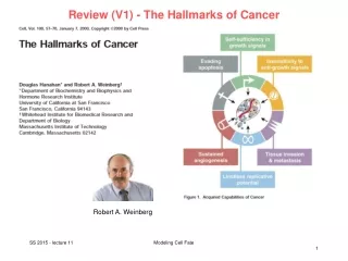
Review (V1) - The Hallmarks of Cancer Robert A. Weinberg Modeling Cell Fate
Review (V1) - The Hallmarks of Cancer Modeling Cell Fate
Review (V1) - Number of somatic mutations in human cancers Top: children vs. adults Numbers in parentheses : median number of nonsynonymous mutations per tumor. MSI, microsatellite instability; SCLC, small cell lung cancers; NSCLC, non–small cell lung cancers; ESCC, esophageal squamous cell carcinomas; MSS, microsatellite stable; EAC, esophageal adenocarcinomas. B Vogelstein et al. Science 2013; 339:1546-1558 Modeling Cell Fate
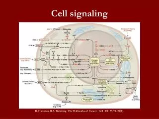
Review (V1) - Cancer driver genes belong to 12 pathways Cancer cell signaling pathways and the cellular processes they regulate. All known driver genes can be classified into one or more of 12 pathways (middle ring) that confer a selective growth advantage (inner circle; see main text). These pathways can themselves be further organized into three core cellular processes (outer ring). B Vogelstein et al. Science 2013; 339:1546-1558 Modeling Cell Fate
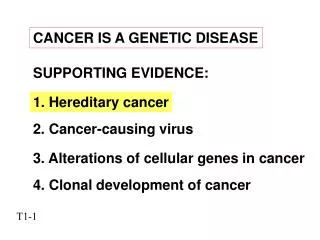
V11 – DNA viruses involved in Cancerogenesis Human papilloma virus (HPV) causes transformation in cells through interfering with tumor suppressor proteins such as p53. Interfering with the action of p53 allows a cell infected with the virus to move into S phase of the cell cycle, enabling the virus genome to be replicated. Some types of HPV increase the risk of, e.g., cervical cancer. Harald zu Hausen Noble price for medicine 2008 www.wikipedia.org Modeling Cell Fate
Epstein-Barr virus The Epstein–Barr virus (EBV), also called human herpesvirus 4 (HHV-4), is a virus of the herpes family, and is one of the most common viruses in humans. Most people on earth become infected with EBV and gain adaptive immunity. EBV infects B cells of the immune system and epithelial cells. While most of the time the infection causes little damage, sometimes the growth activating genes may cause the infected B-cells to turn into cancers in certain people. Epstein-Barr virus is associated with four types of cancers - Post-Transplant Lymphoma and AIDS-Associated Lymphoma - Burkitt's Lymphoma - Hodgkin's Lymphoma - cancer of the nasopharynx (the upper part of the throat behind the nose) The mechanisms how EBV is related to cancerogensis are poorly understood. www.wikipedia.org, lymphoma.about.com Modeling Cell Fate
Computational systems biology of cancer Working hypothesis: Authors propose that viruses and genomic variations alter local and global properties of cellular networks in similar ways to cause pathological states. Study was submitted on June 8, 2011 and accepted only on June 7, 2012! Rozenblatt-Rozen et al. Nature 487, 491 (2012) Modeling Cell Fate
Considered virus ORFs Adenovirus: Nine full length ORFs Epstein-Barr Virus (EBV): Eighty-one EBV ORFs Human Papillomaviruses (HPV): Seven HPV types were chosen for this study: HPV6b, 11, 16, 18 and 33 of the alpha genus, and HPV5 and HPV8 of the beta genus Polyomaviruses: ORF clones were obtained from nine polyomaviruses: BK, HPyV6, HPyV7, JCCY, JCMad1, MCPyV, SV40, TSV and WU. Rozenblatt-Rozen et al. Nature 487, 491 (2012) Modeling Cell Fate
Virome-to-variome network model The virome-to-variome network model proposes that genomic variations (point mutations, amplifications, deletions or translocations) and expression of tumour virus proteins induce related disease states by similarly influencing properties of cellular networks. Rozenblatt-Rozen et al. Nature 487, 491 (2012) Modeling Cell Fate
Virus-host protein-protein interactions (PPIs) Experimental pipeline for identifying virus–host interactions. 123 selected cloned viral ORFs were subjected to yeast 2-hybrid (Y2H) screens against 13000 human ORFs (left), and introduced into IMR-90 lung fibroblast cell lines for both TAP–MS and microarray analyses (right). Numbers of viral ORFs that were successfully processed at each step are indicated in red. Comment: Y2H and TAP-MS are experimental methods to detect physical interactions of 2 (Y2H) or more (TAP-MS) proteins. Rozenblatt-Rozen et al. Nature 487, 491 (2012) Modeling Cell Fate
Binary virus-host PPIs identified by Y2H 31 host target proteins showed more binary interactions with viral proteins (red circles) than would be expected given their ‘degree’ in the current binary map of the human interactome network HI-2. This suggests a set of common mechanisms by which different viral proteins rewire the host interactome network Lines stand for detected protein-protein interactions between viral proteins (open hexagons) and human host proteins (full circles). “Degree” of a protein: number of interaction partners. Rozenblatt-Rozen et al. Nature 487, 491 (2012) Modeling Cell Fate
Enriched GO terms for targeted host proteins With what types of human proteins do viral proteins physically interact? Enrichment of GO terms for host proteins physically interacting with viral proteins. Rozenblatt-Rozen et al. Nature 487, 491 (2012) Modeling Cell Fate
Viral E6 protein Viral E6 protein associates with host E6-AP ubiquitin-protein ligase, and inactivates tumor suppressors TP53 and TP73 by targeting them to the 26S proteasome for degradation. E6/E6AP also degrades other cellular targets including Bak, Fas-associated death domain-containing protein (FADD) and procaspase 8 what causes inhibition of apoptosis. E6 also inhibits immune response by interacting with host IRF3 and TYK2. These interactions prevent IRF3 transcriptional activities and inhibit TYK2-mediated JAK-STAT activation by interferon alpha resulting in inhibition of the interferon signaling pathway. www.uniprot.org Modeling Cell Fate
Specificity of virus-host relationships: PPIs involving E6 • Check protein complex associations mediated by E6 proteins from 6 distinct HPV types representing 3 different disease classes: • high-risk mucosal (dt. (Nasen-)schleim) • low-risk mucosal • cutaneous (dt. kutan, d.h. Haut betreffend) • E6 and E7 proteins encoded by high-risk mucosal HPVs are strongly oncogenic. • Multiple host proteins associate with E6 proteins from 2 or more different HPV types ( P < 0.001). • Transcriptional regulators CREBBP and EP300 only associate with E6 proteins from cutaneous HPV types, but not with those from mucosal classes. • In contrast, no group of host proteins showed class-specific targeting by HCV E7 proteins. Rozenblatt-Rozen et al. Nature 487, 491 (2012) Modeling Cell Fate
Protein complex associations involving E6 proteins Left: Network of protein complex associations of the six E6 viral proteins from 6 HPV types (hexagons, coloured according to disease class) with host proteins (grey circles). Host proteins that associate with 2 or more E6 proteins are colored according to the disease class(es) of the corresponding HPV types. Circle size is proportional to the number of associations between host and viral proteins in the E6 networks. Middle: Distribution plots of 1,000 randomized networks and experimentally observed data (green arrows) for the number of host proteins targeted by 2 or more viral proteins in the corresponding subnetworks. Inset: representative random networks from this distribution. Right: ratio of the probability that a host protein is targeted by viral proteins from the same class to the probability that it is targeted by viral proteins from different classes. Inset: representative random networks from this distribution. Rozenblatt-Rozen et al. Nature 487, 491 (2012) Modeling Cell Fate
Computational systems biology of cancer • Besides targeting protein-protein interactions, viral proteins functionally perturb their hosts through downstream effects on gene expression. • → Profile transcriptome of viral ORF-transduced cell lines to trace pathways through which viral proteins could alter cellular states. • -> 2944 frequently perturbed host genes. • Clustering gives 31 clusters • Many of the clusters are enriched for specific GO terms and KEGG pathways • (p < 0.01) • Identify enriched TF binding motifs in gene promoters or enhancers from data on cell-specific chromatin accessibility and consensus TF-binding motifs. Rozenblatt-Rozen et al. Nature 487, 491 (2012) Modeling Cell Fate
Heatmap of transcriptome perturbations x-axis: 63 microarray experiments where IMR-90 cells were transfected with the indicated viral protein. y-axis: 31 clusters of differentially expressed genes Enriched GO terms and KEGG pathways are listed adjacent to the numbered expression clusters. TFs with enriched binding sites and gene targets enriched for the listed GO and/or KEGG pathways that are physically associated with or differentially expressed in response to viral proteins are shown, with * denoting multiple members of a TF family. Up to 5 TFs are shown for any cluster. Blocks show which viral proteins associate with the indicated host proteins, as detected in our data set (grey) or manually curated (green). Rozenblatt-Rozen et al. Nature 487, 491 (2012) Modeling Cell Fate
Notch pathway Perturbations in Notch signalling can confer either oncogenic or tumour-suppressive effects. Because both inhibition of the Notch pathway and the expression of HPV8 E6 promote squamous cell carcinoma, we reasoned that binding of HPV5 and HPV8 E6 to MAML1 might inhibit Notch signalling. To test this, examine transcript levels of Notch pathway genes and potential Notch target genes with a predicted RBPJ (also known as CSL) binding site in their promoter across all HPV E6 cell lines as well as in cells depleted for MAML1. Rozenblatt-Rozen et al. Nature 487, 491 (2012) www.genome.jp Modeling Cell Fate
Association of HPV E6 proteins with MAML1 inhibits Notch Heat map of expression of Notch-pathway-responsive genes (shown on x-axis from DLL4 to JAG1) in IMR-90 cells on expression of E6 proteins from different HPV types or on knockdown of MAML1, relative to control cells. Transcript levels of several Notch targets were significantly decreased in IMR-90 cells (blue fields) that were either depleted for MAML1 (first line) or expressing either HPV5 or HPV8 E6 (lines 2 and 3). This indicates that the association of HPV5 and HPV8 E6 proteins with MAML1 inhibits Notch signalling. Rozenblatt-Rozen et al. Nature 487, 491 (2012) Modeling Cell Fate
How viral proteins interact with proteins in Notch signaling Representation of viral protein interactions with components of the Notch signalling pathway. Notch ICD, Notch intracellular domain. → viral proteins from all 4 DNA tumour viruses target proteins of the Notch pathway (P < 0.002). This highlights the central role of Notch signalling in both virus–host perturbations and tumorigenesis, and supports observations that implicate MAML1 in cancer pathogenesis. Rozenblatt-Rozen et al. Nature 487, 491 (2012) Modeling Cell Fate
Computational systems biology of cancer To which extent do viral proteins globally target host proteins that have been causally implicated in cancer? Compare the viral targets, identified through binary interactions, protein complex associations and TF-binding-site analyses, against a gold standard set of 107 high-confidence causal human cancer genes in the COSMIC Classic (CC) gene set. Viral targets were significantly enriched among CC genes (P=0.01). Rozenblatt-Rozen et al. Nature 487, 491 (2012) Modeling Cell Fate
Virus-host network model Diagram describing the composition of VirHost (947 proteins identified by TAP–MS with at least 3 unique peptides, Y2H and TF) and overlap with COSMIC Classic (CC) genes. ‘VirHost’ set includes 16 proteins encoded by CC genes (P=0.007), among which tumour suppressor genes were significantly over-represented (P=0.03). Rozenblatt-Rozen et al. Nature 487, 491 (2012) Modeling Cell Fate
Viral proteins, transcription factors and clusters (Left) Network representation of all predicted viral protein-TF-cluster cascades. (Right) Schematic shows how viral protein-TF-target gene network was constructed (Below) representative networks. Rozenblatt-Rozen et al. Nature 487, 491 (2012) Modeling Cell Fate
Compare cancer mutations and PPis with viral proteins Mapping of VirHostSM gene products to both tumours in which they are mutated (left) and to viral interactors (right). Proteins annotated with the GO term “regulation of apoptosis” are indicated in purple. Rozenblatt-Rozen et al. Nature 487, 491 (2012) Modeling Cell Fate
Summary If the mechanisms of cancer formation induced by genetic mutations and by DNA viruses are indeed similar, this opens up interesting possibilities to study cancerogeneis by controlled viral infection. Network view correponds to modern field of cancer systems biology. Important for drug design. Follow-up study which individuals are susceptible to viral infection and which ones are not? Rozenblatt-Rozen et al. Nature 487, 491 (2012) Modeling Cell Fate