Endoscopic Orbital Decompression: A Case Study
480 likes | 597 Views
A 46-year-old female with thyroid orbitopathy presented with right eye pain, underwent decompression surgeries, and received steroid therapy. Follow her progress and treatment plan.

Endoscopic Orbital Decompression: A Case Study
E N D
Presentation Transcript
Case Presentation 12/19 Presenting: clerk 陳豪宏 Instructor: 張丞賢老師
Patient Profile • Name: 謝X莉 • Age: 46 years old • Gender: female • Chart number: 24097375 • Date of admission: 2011/12/12
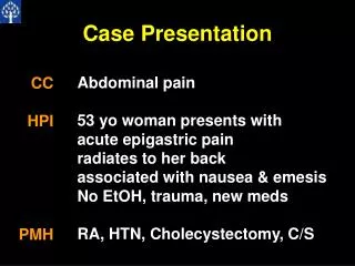
Chief complaint • Right eye pain for 1 week.
Present Illness 1 • This 46 years old female suffered from insidious right eye pain for 1 week. The accompanying symptoms are red eye, tearing (od). She had hyperthyroidism s/p subtotal thyroidectomy on 2011-8-18. • Thyroid orbitopathy with compressive optic neuropathy was diagnosed this September. She was admitted to our hospital and received steroid pulse therapy on September 5th this year.
Present Illness 2 • After discharge on September 9th. She came for OPD follow-up on September 14th. Diplopia was still complained. OCT showed loss of nerve fibrous layer. • She was admitted for operation on September 20th, during which orbital decompression (ou) of temporal sides and muscle recession of inferior rectus were done. • On December 12th, she was admitted for steroid pulse therapy and orbital decompression of nasal sides.
Present Illness Summary First Decompression (Temporal Sides) 09/19~09/23 Thyroidectomy 08/18 First Steroid Therapy 09/05~09/09 Second decompression 12/12~ now(op:12/16)
Past History • hyperthyroidism, s/p operation • hypertension(+) • asthma(+) • heart disease(-) • DM(-) • depressive disorder under medical treatment • Operation history: • L-spine s/p operation 1.5 year ago • hyperthyroidism s/p subtotal thyroidectomy on 2011-8-18 at 謝外科 • Orbital Decompression on 09/20 • Admission history: • As listed in the Present Illness
Personal History • Cigarette Smoking : +, 1/2 pack per day • Alcohol : denied • Occupation history : denied • Contact history : denied • Travel history : denied • Family History: denied family systemic disease • Allergy History: denied
Nutritional and Mental status • Nutritional Status: • Weight: 81.6kg; Height: 151.5cm; BMI: 35.55 • Mental Status: • Consciousness: alert. Mentality: normal.
Current Medicine • 2011-9-13 PSY OPD Dr.葉怡君 • EFEXOR速悅XR(Venlafaxine) 1#AM 1#HS */PC*28 • Estazolam #(Eurodin)管四 #2 QD/HS* 28 • Inderal 10mg (Propranolol) #1 BID/PC* 28 • Xanax 0.25mg (Alprazolam)管四#1 BID/PC* 28 • Zyprexa 5mg (Olanzapine)**** #1 QD/HS* 28
Physical Examination 1 • Vital sign: • BP: 131 / 105 mmHg, PR: 80 bpm, RR: 18 cpm, BT: 37 ℃ • <Ophthalmic examination> 2011/12/12 • OD OS • Eyeball 25>-----------105------------<24 • Conjunctiva injection injection • Cornea clear clear • AC moderate, clear moderate-shallow, clear • Iris np np • Pupil 4mm 4mm • Light reflex +/+ +/+ • Lens clear clear
Physical Examination 2 • Fundus C/D=0.4 C/D=0.7 • disc: temporal pale • EOM 20 20 • | | • 35 ---|---45 45---|---30 • | | • 30 10 • Tonopen: straight: R: 21,22 L: 22,23 • Upward: R: 27,28 L: 27,29 Downward: R: 22 L:22 • Vod: 0.1x 1/5 • Vos: ND/10cm
Orbital CT • Hypertrophy of extraocular muscles (all of ou)
Abnormal Lab finding • 12/12: • Hb: 12.0 Hct: 35.3 • MCV: 79.8 Plt: 502k
Diagnosis • Thyroid orbitopathy with compressive optic neuropathy
Plan: • Pulse therapy of Solumedrol 500mg q12h IV • Arrange Auto-P, VEP, OCT of disc. • Orbital decompression under general anesthesia.
Progress 12/13 • S: eye pain(-) Red eye ↓ • O: EOM: 20 20 40 45 35 10 • 35 (od) 10 (os) • Cornea: clear Conjunctival congestion: ↓ • Chemosis: ↓ AC: deep, clear • A+P: arrange VEP, OCT of disk today. Prepare endoscopic decompression
Progress 12/14 • S: eye pain(-) • O: EOM: 20 20 • 35 45 35 35 35 (od) 10 (os) • Cornea: clear • Conjunctiva: mild congested • Chemosis: ↓ AC: deep, clear • VEP: delayed potency and amplitude (ou) • A+P: Keep pulse therapy day 2
Progress 12/15 • S: Still blurred vision (ou) • O: EOM: 20 20 35 45 40 35 35 (od) 20 (os) • Cornea: clear Conjunctiva: Congestion ↓ ↓ • Chemosis: (-) few SPK in the left lower eye • IOP: 24.7 (os), 20.5 (od). • A+P: Arrange endoscopic orbital decompression (ou) tomorrow
Operation on 12/16 • Endoscopic orbital decompression done in this morning. (8.00 am) • Procedures taken: • Preparation for anesthesia • Endoscope usage: • Use periosteal elevator to destruct cribriform pyparacea temporally and posteriorly. • Destruct the ethmoid bone. • Rigid orbital soft tissue with fibrotic membran was noted. • Incision of the fibrotic membrane and let the orbital fat protrude out from orbital medial wall. • Hemostasis adequately. • The operation was done successfully and the patient returned to the ward for recovery.
Progress 12/16 • S: no obvious pain of wound post-op. Still blurred vision • O: Endoscopic wound of the surgery. Condition fine. • A+P:
Discussion A Brief Introduction of Thyroid Orbitopathy
Thyroid Eye Disease • Thyroid eye disease has many names: • Thyroid-associated orbitopathy (TAO) • Thyroid orbitopathy • Grave's orbitopathy • Among others • All these terms mean the same thing: • inflammation of the tissues around and behind the eye producing varying amounts of swelling and scarring.
Introduction • It is an autoimmune disease. • There are at least various theories to explain its cause and development. • Thyroid eye disease usually occurs in people with a history of thyroid problems. • However, thyroid eye disease can occur decades before, or decades after, the development of thyroid gland disease.
Pathogenesis • The volume of • The extraocular • retroorbital connective and adipose tissue • are increased, due to inflammation and the accumulation of hydrophilic glycosaminoglycans (GAG), principally hyaluronic acid, in these tissues. • GAG secretion by fibroblasts is increased by activated T-cell cytokines such as tumor necrosis factor (TNF) alpha and interferon gamma. • It implies that T-cell activation is an important part of this immunopathology. • The accumulation of GAG causes a change in osmotic pressure, which in turn leads to a fluid accumulation and an increase in pressure within the orbit. • These changes displace the eyeball forward and can also interfere with the function of the extraocular muscles and the venous drainage of the orbits.
Pathogenesis (Summary) • Autoimmune caused Inflammation • T cell: TNF-a and interferon r • Fibroblasts: GAG • Osmotic pressure • Fluid accumulation and IOP • Eyeball forward • Venous drainage malfunction • EOM dysfunction ↑ ↑ ↑ ↑
Risk Factors • Treatment: Ra-I therapy may be more likely to lead to the development or worsening of TO than medication or subtotal thyroidectomy. • Sex: TO: female > male • According to studies, smoking definitely makes thyroid eye disease worse. • increase in the connective tissue volume of the orbit, but not the extraocular muscle volumes. • Thyrotropin receptor autoantibodies • Higher titer -> Higher prevalence and longer course of TO.
Epidemiology • Approximately 20 to 25 percent of patients with Graves' hyperthyroidism have clinically obvious TO. • Along with the eye signs of thyroid hormone excess (lid retraction and stare), at the time of diagnosis of the hyperthyroidism. • However, many more patients with Graves' hyperthyroidism have evidence of TO in imaging studies.
Clinical Presentations • proptosis and periorbital edema
Clinical Presentations 2 • The patient may be distressed by the appearance of his or her eyes. • Possible major symptoms: • sense of irritation • excessive tearing that is often made worse by exposure to cold air, wind, or bright lights • eye or retroorbital discomfort or pain • blurred vision • Diplopia • and occasionally loss of vision.
Proptosis • The degree of proptosis (exophthalmos) is dependent on the depth of the orbit and the degree of enlargement of the retroocular muscles and retroorbital fibrous and fatty tissue. • The proptosis may be usually symmetric, but is often asymmetric, and may be accompanied by a sensation of pressure behind the eyeballs. • The proptosis may be masked by periorbital edema, which is a common accompaniment.
Apparent proptosis • Many patients with hyperthyroidism have lid retraction secondary to thyroid hormone excess. • It leads to stare and lid lag, resulting from contraction of the levator palpebrae muscles of the eyelids. • The stare may give the appearance of proptosis, when none in fact exists ("apparent proptosis"). • These signs alone do not indicate the presence of ophthalmopathy, and subside when the hyperthyroidism is treated.
Physical Examinations 1 • Inspection of the conjunctivae and periorbital tissue, looking for chemosis and periorbital edema. • Determination of the extent to which the upper and lower lids can be closed. • Because failure of apposition promotes drying and ulceration of the cornea. • Assessment of EOM. • inability to achieve or maintain convergence. • Limitation of upward gaze. It leads to a characteristic head-back position in order to see ahead. • double vision.
Physical Examinations 2 • exophthalmometer. • measurement of the distance between the lateral angle of the bony orbit and an imaginary line tangent to the most anterior part of the cornea. • The upper limit of normal is 20~22 mm. • as high as 30 mm in patients with severe proptosis. • Visual acuity and color vision should be assessed by simple reading tests and color charts, and visual fields should be evaluated by confrontation.
Assessment of Severity • NO SPECS by American Thyroid Association • Class 0 — No symptoms or signs • Class I — Only signs, no symptoms (eg, lid retraction, stare, lid lag) • Class II — Soft tissue involvement • Class III — Proptosis • Class IV — Extraocular muscle involvement • Class V — Corneal involvement • Class VI — Sight loss (optic nerve involvement)
Differential Diagnosis • eye signs of thyroid hormone excess • Bilateral eye signs simulating TO: • severe obesity • Cushing's syndrome • orbital myositis • histiocytosis • myasthenia gravis • very rarely: orbital tumors • statin-induced EOM myopathy.
Differential Diagnosis 2 • For possible unilateral TO, space-occupying lesions of the orbit must be ruled out first. • When necessary, the diagnosis can be confirmed by ultrasonography and CT. • It is important not to inject iodinated contrast material in patients with Graves' disease especially if radioiodine therapy is contemplated. • If diagnos is not in doubt, only tests necessary are: • serum TSH • Free T4 • TSHR antibodies.
Treatment • treat according to the severity. • Most patients have mild disease and do not have progression during follow-up. • The treatment of TO includes • reversal of hyperthyroidism • relief of symptoms • reduction of inflammation in the periorbital tissues.
Effects of anti-hyperthyroidism therapies • Subtotal thyroidectomy and antithyroid drugs do not appear to have a negative influence on the course of orbitopathy. • However, there is increasing evidence that radioiodine therapy can cause the development or worsening of Graves' orbitopathy more often than antithyroid drug therapy or surgery.
Symptomatic treatment • Eye shades • Artificial tears (saline eye drops) • Raising the head of the bed when sleep. • Photophobia and sensitivity to wind or cold air can be relieved by use of dark glasses. • Glucocorticoids (oral or IV), are the primary treatment for severe Grave‘s orbitopathy. • Radiation and surgical decompression can also be used in selected patients.
Therapies for severe TO • Oral Prednisone: • effective treatment for TO. More side effects. It has been seen to induce liver failure. • Intravenous glucocorticoid pulse therapy: • Fewer side effects and better clinical outcome in • Radiotherapy: benefits controversial
Orbital decompression surgery • Three major indications: • If glucocorticoid therapy or orbital irradiation fails to halt progression of TO • If loss of vision is threatened either by • ulceration or infection of the cornea • changes in the retina or optic nerve • For cosmetic correction of severe proptosis • surgery should be avoided for as long as possible until the disease stabilizes under corticosteroid suppression
Orbital decompression surgery • The orbit may be decompressed by removing the lateral wall, the roof, or the medial wall and the floor. • Uptodate suggests the last procedure, also known as transantral decompression. • the surgeon removes the floor and medial wall of the orbit to allow decompression. • It does not leave a scar on the face, and avoids craniotomy.
Result of decompression surgery • An excellent result can usually be achieved, with substantial reduction in proptosis and edema. • However, diplopia usually does not improve and may worsen, so that eye muscle surgery is almost always needed later. • Timing of Surgery: Clinical outcome appears to be better if decompression surgery is performed after rather than before glucocorticoid therapy.
Other operations • Fat decompression surgery: • Removal of the retroorbital adipose tissue • Bilateral lateral tarsorrhaphy may be performed to minimize or prevent corneal damage. • Surgical recession of Muller's muscle and the levator will correct upper lid retraction. • However, decompression surgery is preferable for both of these problems because it is more effective both functionally and cosmetically.
Reference • Uptodate articles: • Pathogenesis and clinical features of Graves' ophthalmopathy (orbitopathy) • Treatment of Graves' orbitopathy (ophthalmopathy)
The End • Thank you.