Download
1 / 35
350 likes | 524 Views
Block: URIN 313 Physiology of THE URINARY SYSTEM Lecture 1. Dr. Amel Eassawi Dr. Shaikh Mujeeb Ahmed. STRUCTURE AND FUNCTIONS OF THE NEPHRON . OBJECTIVES. The student should be able to: List and understand the major functions of the kidneys.

E N D
Block: URIN 313Physiology of THE URINARY SYSTEM Lecture 1 • Dr. AmelEassawi • Dr. ShaikhMujeeb Ahmed
OBJECTIVES • The student should be able to: • List and understand the major functions of the kidneys. • State the role of erythropoietin and the stimulus for its secretion • Describe the location of the kidneys and their gross anatomical features. • Describe the different parts of the nephron and their location within the kidney. • List the individual nephron segments, in order and identify each structure as being in the cortex or medulla. • List the structures of the renal circulation and their association with different aspects of the nephron. • Distinguish between superficial and juxtamedullarynephrons. • Describe the essential elements of the juxtaglomerular apparatus and its function in the nephron. • Describe the basic renal process of urine formation.
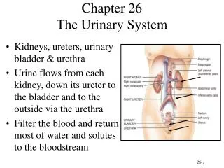
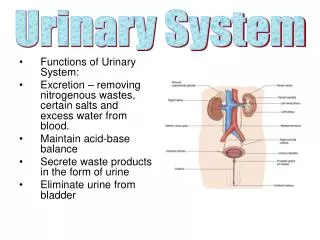
Urinary System • Consists of • Urine forming organs • kidneys • Structures that carry urine from the kidneys to the outside for elimination from the body • Ureters • Urinary bladder • Urethra
Kidney Functions • Urine formation • Maintain H2O balance in the body • Maintain osmolarityof body fluids, primarily through regulating H2O balance • Regulate the quantity and concentration of most ECF ions e.g. Na+, Cl-, K+, Ca2+, H+, HCO3-, PO43- • Maintain plasma volume • Help maintain acid-base balance in the body: • Excrete acids (kidneys are the only means of excreting non-volatile acids, such as sulfuric acid and phosphoric acid). 2. Regulate body fluid buffers ( e.g. Bicarbonate).
Kidney Functions • Excreting (eliminating) the end products (wastes) of bodily metabolism e.g. urea (from protein metabolism), uric acid (from nucleic acid metabolism), creatinine(from muscle metabolism), bilirubin(from hemoglobin metabolism). • Excreting metabolites of various hormones. • Excreting - drugs, food additive. • Produce erythropoietin. • Produce Renin. • Convert vitamin D into its active form. • 1,25 dihydroxycholecalciferol • In vivo vitamin D synthesis involves a series of biochemical transformations, the last of which occurs in the kidneys. The active form of vitamin D (1,25-dihydroxyvitamin D3) ( also known as calcitriol), is activated in the kidneys, and its rate of synthesis is regulated by hormones that control calcium and phosphate balance.
Regulation of Erythrocyte Production Imbalance Start Normal blood oxygen levels Stimulus: Hypoxia due to decreased RBC count, decreased availability of O2 to blood, or increased tissue demands for O2 Imbalance Increases O2-carrying ability of blood Reduces O2 levels in blood Enhanced erythropoiesis increases RBC count Erythropoietin stimulates red bone marrow Kidney (and liver to a smaller extent) releases erythropoietin Kidneys produce Erythropoietin: a hormone that stimulates bone marrow to produce red cells.
Renin-Angiotensin-Aldosterone System Renin is an enzyme secreted by the kidneys from granular cells of the juxtaglomerularapparatus. It activates the renin-angiotensin system by converting angiotensinogen to angiotensin I.
Kidneys • The kidneys are a pair of bean-shaped organs that lie at the back of abdominal cavity. • Kidneys are retroperitoneal (behind the peritoneum). • Each kidney is about 4-5 inches (10-12 cm) in length. • Each kidney is supplied with a renal artery and a renal vein • Acts on plasma, flowing through it to produce urine • Formed urine drains into the renal pelvis, Located at medial inner core of each kidney • Urine is drained into ureters, and stored temporarily in the urinary bladder before it’s emptied through the urethra to outside the body.
Ureters • Smooth muscle-walled duct. • Exits each kidney at the medial border in close proximity to renal artery and vein. • Carry urine to the urinary bladder.
Urinary Bladder • Temporarily stores urine. • Hollow, distensible, smooth muscle-walled sac. • Periodically empties to the outside of the body through the urethra.
Urethra • Conveys urine to the outside of the body • In females Urethra is straight and short about 4cm long • In males • Much longer about 19- 20cm and follows curving course from bladder to outside • Has dual function • Provides route for eliminating urine from bladder. • Passageway for semen from reproductive organs. • In male, Prostate gland lies below the neck of bladder and completely encircles the urethra. • Prostate gland enlargement occurs during middle to older age and can occlude (block) the urethra, therefore, obstructing the flow of urine.
Nephron • Functional and structural unit of the kidney • Approximately 1 million nephrons/kidney • Each nephron has two components • Vascular component • Tubular component • Arrangement of the nephrons within the kidney gives rise to two distinct regions • Outer cortex Renal cortex (granular in appearance) • Inner medulla Renal medulla -made up of striated triangles called renal pyramids
Vascular component of the nephron • The renal corpuscle consists of a compact tuft of interconnected capillary loops, the glomerulus (pl. glomeruli) or glomerular capillaries, surrounded by a balloon-like hollow capsule: Bowman’s capsule. Blood enters and leaves Bowman’s capsule through arterioles that penetrate the surface of the capsule at the vascular pole.
Vascular component of the nephron • Renal Artery enters the kidney and forms afferent arteriole, which supplies each nephron. • Afferent arteriole delivers blood to the glomerulus. • Glomerular capillaries rejoin to form another arteriole – the efferent arteriole. • The blood that was not filtered in the glomerulus goes to efferent arteriole. • Efferent arteriole gives second set of capillaries, the peritubular capillaries, which supply the renal tissue and are important in exchange with tubular system. (Peritubular means around the tubular system) • Peritubular capillaries rejoin to form venules, that drain in renal vein.
Tubular component of the nephron • Hollow, fluid-filled tube • single layer of epithelial cells • Components • Bowman’s capsule • Proximal convoluted tubule • Loop of Henle • Descending limb (thin) • Ascending limb (thin and thick part) • Distal convoluted tubule • Collecting duct or tubule • Juxtaglomerular apparatus
Tubular component of the nephron • Bowman’s capsule – expanded double walled invagination that cups around the glomerulus to collect fluid from the glomerular capillaries. • From bowman’s capsule, filtered fluid passes into PCT. PCT lies entirely in the cortex. • From PCT, fluid passes into loop of Henle [LH]. • Loop of Henle – form U-shaped or hair pin loop, LH dips into renal medulla.
Tubular component of the nephron • Loop of Henle – descending limb of LH goes from cortex to medulla, and ascending limb of LH passes from medulla to cortex. • Ascending limb of LH, forms distal convoluted tubule (DCT), DCT lies in cortex. • Distal convoluted tubule DCT empties into the collecting tubule or duct. • Collecting tubule gets fluid from about 8 nephron (DCT). • Collecting tubule passes from cortex to medulla and empties into renal pelvis.
Juxtaglomerular apparatus • Distal convoluted tubule, afferent and efferent arterioles are specialized to form Juxtaglomerular apparatus (JGA). • Cells in the DCT are called Macula densa and the cells in the afferent (mainly) and efferent arteriole which contain secretary cells are called Juxtaglomerular cells. • The combination of Macula Densa and Juxtaglomerular cells is called Juxtaglomerular apparatus. • Juxtaglomerular apparatus secretes Renin in blood.
Nephron Two Types of Nephrons • Distinguished by location and length of their structures • Juxtamedullary nephrons • Cortical nephrons
Cortical nephrons • - About 80% nephron • have glomeruli located in the outer cortex. • have short loops of Henle that penetrate only a short distance into the medulla, • In cortical nephron, peritubular capillaries do not form vasarecta, but go around the short loop of Henle • Involved in solute reabsorption.
Juxtamedullary nephrons • About 20 per cent of the nephrons • have glomeruli that lie in the inner layer of the renal cortex near the medulla • have long loops of Henle that dip deeply into the medulla. • - have peritubular capillaries called vasarecta (straight vessels) which run with the loop of Henle • - Play important role in concentration mechanism of urine
IMPORTANT • All nephron originate in the cortex. • Glomeruli of cortical nephron lie in the outer layer of cortex and glomeruli of Juxta medullary nephron lie in the inner layer of the cortex near the medulla • The kidney cannot regenerate new nephrons. • Aging causes a gradual decrease in nephron number.
URINE FORMATION Basic Renal Processes • Glomerularfiltration • Tubular reabsorption • Tubular secretion Urine results from these three processes. Excretion = Filtration – Reabsorption + Secretion
REFERENCES • Human Physiology by Lauralee Sherwood, Seventh edition • Text Book of Physiology by Guyton &Hall,11th edition • Text Book of Physiology by Linda .s Contanzo, Third edition