Background
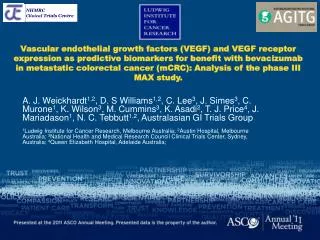
Vascular endothelial growth factors (VEGF) and VEGF receptor expression as predictive biomarkers for benefit with bevacizumab in metastatic colorectal cancer (mCRC): Analysis of the phase III MAX study.

Background
E N D
Presentation Transcript
Vascular endothelial growth factors (VEGF) and VEGF receptor expression as predictive biomarkers for benefit with bevacizumab in metastatic colorectal cancer (mCRC): Analysis of the phase III MAX study. A. J. Weickhardt1,2, D. S Williams1,2, C. Lee3, J. Simes3, C. Murone1, K. Wilson3, M. Cummins3, K. Asadi2, T. J. Price4, J. Mariadason1, N. C. Tebbutt1,2, Australasian GI Trials Group 1Ludwig Institute for Cancer Research, Melbourne Australia; 2Austin Hospital, Melbourne Australia; 3National Health and Medical Research Council Clinical Trials Center, Sydney, Australia; 4Queen Elizabeth Hospital, Adelaide Australia;
Background • To date there is no reliable, validated predictive biomarker for bevacizumab benefit in metastatic colorectal cancer (mCRC)1 • The potential role of alternate VEGFR-2 ligands such as VEGF-C and VEGF-D as predictors of bevacizumab benefit was previously untested • VEGF-C and VEGF-D have been demonstrated in in vitro studies as capable of stimulating angiogenesis through binding to VEGFR22,3 Bevacizumab VEGF-A • Jubb et al, Lancet Oncology, 2010. • Achen et al, Int J Exp Path, 1998. • Rissanen et al, Circ Res, 2003
Background • Plausibly, these ligands could continue to stimulate angiogenesis even if bevacizumab abrogated signaling via VEGF-A. High expression of these ligands could constitute a mechanism of resistance to bevacizumab4 • We undertook an analysis of the expression of VEGF family members (VEGF A-D) as well as VEGFR1 and VEGFR2 in tumour tissue by immunohistochemistry (IHC) • We evaluated their role as predictors of efficacy of bevacizumab by correlating expression with clinical outcomes observed in the MAX study 4. Jubb et al, Clin Cancer Res, 2011
Background • The AGITG MAX (Mitomycin, Avastin and Xeloda) study5 was an investigator led study evaluating the impact on progression free survival (PFS) of the addition of bevacizumab to capecitabine chemotherapy in first line therapy for metastatic colorectal cancer (mCRC). • 471 patients were randomised to receive either • capecitabine (C) • capecitabine and bevacizumab (CB) • capecitabine, bevacizumab and mitomycin (CBM) • The results have previously been reported5, demonstrating an improvement in PFS with the addition of bevacizumab, but no improvement in overall survival (OS) 5. Tebbutt N et al. J Clinical Oncology, 2010
Methods • Formalin fixed, paraffin embedded samples of patient tumour tissue were retrieved from patients participating in the biomarker sub-study • Tissue Microarrays (TMAs) were constructed using 1mm tumour cores (triplicates) • Renal cortex cores used as positive controls for staining in the TMAs • IHC analysis took place using standard procedures • Slides created from 4μm sections from the TMA were deparaffinised, washed, then underwent antigen retrieval, exposure to the primary antibody, secondary antibody, and monitored for development of any signal. • Concordance between independent scorers for IHC (weighted Kappa) was >0.79 • Primary tumour used for IHC expression 83%
Methods Kidney 3+ 2+ 1+ 0 Distribution scoring VEGF-A • Scoring • performed independently by two scorers • blinded to treatment allocation and outcome • staining determined to be 0, 1+, 2+ or 3+, depending on intensity of staining. • focal staining of any intensity when only <25% of tumour cells stained was scored as 1+. • The mean score from triplicates was used • Discrepancies between scorers resolved by consensus on second review. VEGF-B VEGF-C VEGF -D VEGF-R1 VEGF-R2
Statistical analysis • A protocol for statistical analysis was developed blinded to knowledge of treatment allocation and patient outcomes. • The choice of cut points for each biomarker (0,1 vs 2 vs 3) was based on pooled distribution of all three treatment groups. • The primary endpoint was PFS, OS was the secondary endpoint. • Each of the biomarkers was initially examined singly. • Kaplan-Meier curves were used to summarise survival outcomes according to treatment, stratified by biomarker expression. • Proportional hazards model with treatment covariates (C vs CB and CBM), biomarker expression, and their interaction was used to assess whether increasing biomarker expression was predictive of resistance to bevacizumab. • Multivariate adjustments for baseline clinical/pathological factors performed and all other six biomarkers were also performed. • The majority of patients (57%, 268/471) had tumour biomarker testing performed
Statistical analysis Consort Diagram: MAX and Biomarker sub-study Randomly assigned (n = 471) Allocated to intervention CBM (n=158) • Allocated to intervention C (n=156) • Withdrew consent (n=1) • Refused treatment (n=1) • Allocated to intervention CB (n=157) • Died (n=1) Received allocated intervention (n=154) Received allocated intervention (n=156) Received allocated intervention (n=158) Did not consent to additional testing (n=31) Tissue specimen insufficient or unavailable (n=37) Did not consent to additional testing (n=24) Tissue specimen insufficient or unavailable (n=41) Did not consent to additional testing (n=27) Tissue specimen insufficient or unavailable (n=43) Tissue specimens tested for biomarkers (n=88) Not evaluable for biomarkers (n=3) Tissue specimens tested for biomarkers (n=92) Not evaluable for biomarkers (n=5) Tissue specimens tested for biomarkers (n=88) Not evaluable for biomarkers (n=5)
Statistical analysis • Biomarker population was representative of MAX ITT population
Results VEGF-D 0,1+ • PFS: low expression of VEGF-D is predictive for bevacizumab benefits • The additional benefit of bevacizumab on PFS was significantly greater among the patients with lower expression of VEGF-D those with higher expression of VEGF-D (interaction p =.02). • Results remained significant after adjustment for baseline clinical/pathological factors (interaction p =.04) and other biomarkers (interaction p=.04). Global test of biomarker-treatment interaction is not significant (p=.22). VEGF-D 2+ VEGF-D 3+
Results VEGF-D 0,1+ • OS: low expression of VEGF-D is predictive for bevacizumab benefits • The additional benefit of bevacizumab on OS was significantly greater among patients with lower expression of VEGF-D than those with higher expression of VEGF-D (interaction p=.01). • Results remained significant after adjustment for baseline clinical/pathological factors (interaction p=.02) but not with other biomarkers (interaction p=.24). VEGF-D 2+ VEGF-D 3+
Results PFS: low expression of VEGF-D is predictive for bevacizumab benefits OS: low expression of VEGF-D is predictive for bevacizumab benefits FOREST PLOT: PROGRESSION FREE SURVIVAL AND VEGF-D HR (95% CI) p-interaction 0.21 (0.08 to 0.55) VEGF-D 0, 1+ 32 0.67 (0.45 to 1.00) VEGF-D 2+ 117 0.02 0.77 (0.50 to 1.17) VEGF-D 3+ 110 0.61 (0.50 to 0.74) C vs CB + CBM 471 0.2 0.4 0.6 0.8 1.0 1.2 CB + CBM better C better
Results P-values indicate the level of significance for the interaction between treatment (C vs CB + CBM) and biomarker for the unadjusted (*), adjusted for baseline clinic-pathologic characteristics (#), and adjusted for other biomarkers (†)
Results PFS: low expression of VEGF-D is predictive for bevacizumab benefits • The additional benefit of bevacizumab on PFS was significantly greater among the patients with lower expression of VEGF-D than those with higher expression of VEGF-D (interaction p =.02). • Results remained significant after adjustment for baseline clinical/pathological factors (interaction p =.04) and other biomarkers (interaction p=.04). Global test of biomarker-treatment interaction is not significant (p=.22). • VEGF-D expression did not appear prognostic for PFS in the capecitabine alone arm • however study underpowered to answer this definitively OS: low expression of VEGF-D is predictive for bevacizumab benefits • The additional benefit of bevacizumab on OS was significantly greater among patients with lower expression of VEGF-D than those with higher expression of VEGF-D (interaction p=.01). • Results remained significant after adjustment for baseline clinical/pathological factors (interaction p=.02) but not with other biomarkers (interaction p=.24).
Results Other results: low VEGFR-1 predictive for bevacizumab benefit on OS • Global test of biomarker-treatment interaction for OS is significant (p=.02). • In a step down multivariable analysis with adjustment for treatment biomarker interactions, then only VEGFR-1 interaction remained significant (p=.02). • Significance of under expression of VEGFR-1 and enhanced benefit of bevacizumab on OS is uncertain because: • outcome limited to OS but not PFS. • no clear biological explanation and not supported by other studies6. 6. Foernzler, D et al, ASCO Gastrointestinal Cancer Symposium, 2010
Discussion • This is a large biomarker study based on a phase III trial of bevacizumab in mCRC • Given VEGF-C and VEGF-D can bind to VEGFR-2 and lead to angiogenesis, we hypothesised that over expression would lead to a decrease in effectiveness of bevacizumab, which would only block VEGF-A interaction with the receptor. • IHC analysis was performed for VEGF A-D, VEGFR-1 and VEGFR-2 expression • No previously validated scoring system for IHC, so used simplified system • Two independent scorers, with good inter-rater agreement • Majority of tumour samples from primary cancer (83%). The correlation of expression of angiogenic factors between primary and metastases is unknown. However the use of the primary reflects the current clinical practice of not performing rebiopsy in most mCRC patients.
Discussion VEGF-D expression is a promising predictive biomarker for bevacizumab • VEGF-D under expression corresponded with enhanced benefit from the addition of bevacizumab to chemotherapy in mCRC. • Consistent statistical significance in analysis of both PFS and OS. • Significance retained in step-down multivariable analysis for PFS. • Strengths of this study are that results are based on randomised comparison from a cohort of patients with same treatment effects as bevacizumab as for the full trial, methods of scoring and analysis done blinded to knowledge to treatment effects by biomarker status. • Limitations are that while findings for VEGF-D are significant for PFS, they do involve small numbers in the VEGF-D 0-1 group. The findings are not as clear for OS, where VEGFR-1 appeared more significant. Also a global test for interaction for PFS did not reach statistical significance, indicating the results should still be regarded as exploratory.
Conclusions Implications and future directions • Further confirmatory studies are required before clinical implementation of IHC expression levels of VEGF-D use as predictive biomarker of bevacizumab.This will include use of an alternate anti-VEGF-D antibody and testing of samples where patients received bevacizumab with different chemotherapy regimens. • If confirmed, there are two significant implications • Bevacizumab use could be restricted to patients with VEGF-D under expression, enhancing clinical benefit and reducing clinical cost burden • VEGF-D could serve as a therapeutic target either alone or in combination with bevacizumab to inhibit angiogenesis and enhance chemotherapy efficacy