Magnetic Field (B)
150 likes | 386 Views
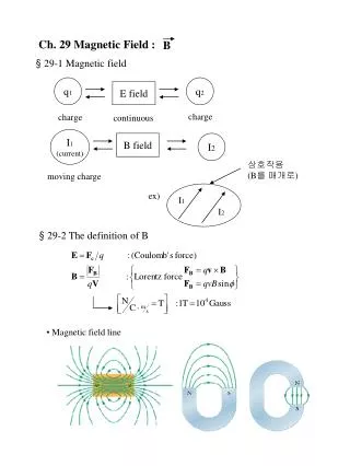
Magnetic Field (B). A photon generates both an electric and a magnetic field. A current passing through a wire also generates both an electric and a magnetic field. . B (T) = |B| = m o I /(2 p r) permittivity of vacuum: m o = 4 p x 10 -7 T m amp -1 I = current (amp)

Magnetic Field (B)
E N D
Presentation Transcript
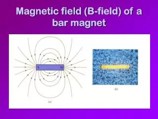
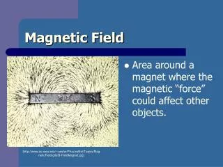
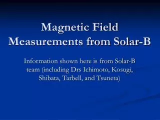
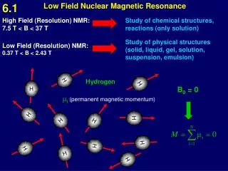
Magnetic Field (B) A photon generates both an electric and a magnetic field A current passing through a wire also generates both an electric and a magnetic field B (T) = |B| = moI/(2pr) permittivity of vacuum: mo = 4p x 10-7 T m amp-1 I = current (amp) r = distance from wire (m) Magnetic Spectroscopy Zeeman Spectroscopy: A magnetic field will eliminate degeneracy of various mℓ quantum states. e.g. An 1S → 1P transition will be split into 3 bands. ESR Spectroscopy: (microwave region) Applies to an molecule with unpaired electrons (e.g. radicals & TM cpds). Hold frequency constant and vary B. Position of transition depends on interactions with adjacent nuclear spins.
Zeeman Spectroscopy Look at the 1S → 1P electronic transition in an atom (e.g. He 1s → 2P transition) ℓ = 0 to ℓ = 1 L = 1 and ML = -1, 0, +1 (split) L = 1 and ML = -1, 0, +1 (degenerate) ←ML= +1 ML = 0 → ← ML= -1 1P 1P DE DE L = 0 and ML = 0 1S 1S no magnetic field With magnetic field
ESR Spectroscopy (microwave region) ΔE = geμBB0 ge = 2.0023 B ≠ 0 B = 0
Proton NMR Spectroscopy Spin (I) = ½ Possible orientations = 2I + 1 = 2 DEmag = gN • mN • B • MI gN = unitless constant related to magnetic moment = 5.586 for proton mN = nuclear magneton B = magnetic field strength MI = nuclear spin angular momentum quantum # Btot = B (1- s) s = shielding constant d = chemical shift d (ppm) = (sTMS – snuc) x 106
Proton NMR Spectroscopy Spin (I) = ½ Possible orientations = 2I + 1 = 2 DEmag = gN • mN • B • MI Btot = B (1- s) s = shielding constant d = chemical shift d (ppm) = (sTMS – snuc) x 106 spin-spin coupling – splits absorption into multiple values like Zeeman selection rules do not allow spin-spin coupling of Hs on same C atom … but do allow coupling of Hs on adjacent Cs. example 16.12: methane – ethane - propane doublet = 1:1 triplet = 1:2:1 quadruplet = 1:3:3:1
NMR Spectroscopy: (radio wave region) Applies to nuclei that have a total spin (I) Application of a magnetic field splits energy levels according to MI values. Position of transition depends on interactions with adjacent nuclear spins. DB = -sBs = shielding constant (no units) Bo is constant and vary frequency Btot = Bo (1 – s) hν = gNμNB0
DE = mB/I m = magnetic moment like dipole moment for magnetic rather than charge I = spin
Pulsed Fourier Transform Spectroscopy Relaxation time (T1)
Relaxation Time T1 = spin-Lattice Relaxation or longitudinal relaxation The time it takes for a fraction of the spins to re-equilibrate after a pulse. Sample is ready to be pulsed again. Relaxation Time T2 = transverse relaxation The time it takes for pulsed nuclei precessing in sync to fall out of synchronization. T2 is typically shorter that T1 and related to linewidths of NMR lines.
1D NMR – magnetic field is constant – vary emrn, at E = hn = DE the nucleus will absorb emr resulting in NMR spectra. The chemical shift (relative value of n) and splitting pattern is dependent on the chemical environment of the proton. 2D NMR – Main magnetic field is constant (B0) A 2nd magnetic field (B1) ┴ to (B0) Relaxation time – fraction of time required after pulse for nuclear spins to return to their original equilibrium distribution values. Boltzman Distribution Function N2/N1 = e-DE/kT. NOE – Nuclear Overhauser Effect – Through space interactions that influence the relaxation time of neighboring protons. Used for 2D NMR structure determination of proteins.
fMRI A techniques that monitors the NMR signal of water in a specific area of the brain – 64 MHz with 1.5T instrument The water signal is influenced by the Fe center of Hemoglobin deoyHb is paramagnetic and shifts the water signal. Since the signal is spread out over a greater n range the spectrum records this as a reduction in signal intensity relative to oxyHb.
BOLD Blood Oxygen Flow Dependence Baseline brain activity = normal blood flow Active areas of brain ↑metabolic activity This causes ↑glucose metabolism and ↑O2 release and ↑[deoxyHb] Body responds by increasing blood flow to area for 4-5s – mechanism ? ↑blood volume = ↑(oxyHb/deoxyHb) = ↑(H2O NMR signal)
Control – no cognitive activity NMR signal intensity Noise or normal fluctuations in signal intensity time Increase in oxyHb due to blood flow (BOLD) Blood Oxygen Flow Dependence NMR signal intensity 4-5s Increase in deoxyHb Cognitive activity in specific brain area time