Download

1 / 81
880 likes | 1.28k Views
Knee MRI Fall 90. Anterior Cruciate Ligaments (ACL). In extension with the medial femoral condyle removed depicts laxity of the anteromedial bundle (red) and a taut posterolateral band (blue). In flexion, the anteromedial band (red) becomes taut and the posterolateral band (blue) becomes lax.

E N D
In extension with the medial femoral condyle removed depicts laxity of the anteromedial bundle (red) and a taut posterolateral band (blue). In flexion, the anteromedial band (red) becomes taut and the posterolateral band (blue) becomes lax.
Kissing anterior bone bruises indicative of hyperextension mechanism in a patient with an ACL tear (ACL not shown). Sagittal T1-weighted MRI shows apposing ill-defined hypointense bone contusions of the anterior femur and adjacent tibial plateau. T2-weighted imaging is more sensitive than T1-weighted imaging for detecting acute bone bruises. A small, linear incomplete subchondral fracture is superimposed on the tibial bone bruise.
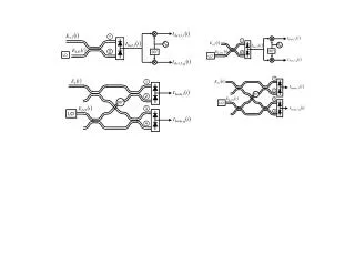
Prescribing sagittal images. Images are obtained no more than 10° oblique to a perpendicular to a line connecting the posterior femoral condyles (the bicondylar line). Alignment of sections directly along the long axis of the anterior cruciate ligament (ACL) in the axial plane is discouraged; this will often lead to overly oblique sagittal images with degraded visualization of the menisci and other knee structures.
Nonvisualization of the ACL as a primary sign of a tear in a T2-weighted sequence. Sagittal T2-weighted fat-saturated fast spin-echo (FSE) image shows replacement of the ACL in the intercondylar notch by extensive edema-fluid signal intensity.
Posterior cruciate ligament (PCL) tear in a patient with a hyperextension-dislocation injury and an anterior cruciate ligament (ACL) tear. Sagittal T1-weighted MRI shows an enlarged, hypointense (obviously torn) PCL (arrow).
(Same patient as in previous image.) Acute hyperextension-dislocation tear of the ACL. Sagittal T1-weighted image shows ill-defined edema/hemorrhage in the intercondylar notch in the usual location of the ACL; no normal ACL fibers are identified. This is a common appearance of an acute ACL tear on MRI images. Some combination of ACL, posterior cruciate ligament, and lateral collateral ligament tears are often present with hyperextension-dislocation injuries of the knee.
Contrecoup bone bruise of the tibia in a patient with an ACL tear. High-signal bone bruise in the posteromedial aspect of the tibial plateau (arrow). This is a common finding in patients with ACL tears secondary to a pivot-shift/twisting mechanisms. This bone bruise is a marker for a pronounced twisting injury. As such, meniscal tears are exceptionally frequent. (Even when no meniscal tears are seen, occult medial meniscal-meniscocapsular junction tears are not uncommonly present.)
Acute tear of the ACL manifest as focal interruption of the ligament. T2-weighted image shows focal hyperintense edema and/or fluid involving the proximal ACL.
Kissing anterior bone bruises indicative of hyperextension mechanism in a patient with an ACL tear (ACL not shown). Sagittal T1-weighted MRI shows apposing ill-defined hypointense bone contusions of the anterior femur and adjacent tibial plateau. T2-weighted imaging is more sensitive than T1-weighted imaging for detecting acute bone bruises. A small, linear incomplete subchondral fracture is superimposed on the tibial bone bruise.
Posterior cruciate ligament (PCL) tear in a patient with a hyperextension-dislocation injury and an anterior cruciate ligament (ACL) tear. Sagittal T1-weighted MRI shows an enlarged, hypointense (obviously torn) PCL (arrow).
(Same patient as in previous image.) Acute hyperextension-dislocation tear of the ACL. Sagittal T1-weighted image shows ill-defined edema/hemorrhage in the intercondylar notch in the usual location of the ACL; no normal ACL fibers are identified. This is a common appearance of an acute ACL tear on MRI images. Some combination of ACL, posterior cruciate ligament, and lateral collateral ligament tears are often present with hyperextension-dislocation injuries of the knee.
(Same patient as in previous image.) Fibular collateral ligament tear in a patient with a hyperextension-dislocation injury. Coronal T1-weighted MRI shows a torn, displaced fibular collateral ligament (FCL) (arrow). The fibular collateral ligament is a major component of the lateral collateral ligament complex. Some combination of lateral collateral ligament, anterior cruciate ligament, and posterior cruciate ligament tears are often present in patients with hyperextension injuries to the knee.
Pivot shift bone bruises of the femur and tibia as a secondary sign of an ACL tear. Sagittal T2-weighted fast spin-echo fat-saturated MRI shows typical pivot-shift subchondral bone bruises of the posterior lateral tibial plateau and lateral femoral condyle near the anterior horn meniscus. The probability of an ACL tear is quite high if both bone bruises are present, only slightly lower if the tibial bone bruise is present in isolation, and only slightly lower still with an isolated femoral bone bruise of this appearance.
Pivot-shift osteochondral fracture of the lateral femoral condyle. This is a sagittal gradient-echo MRI in 17-year-old boy with an arthroscopically proven ACL tear. The fracture is manifest by focal cortical indentation of lateral femoral condyle (arrow), near the anterior horn lateral meniscus. This fracture was accompanied by typical femoral and tibial pivot-shift bone bruises, as best demonstrated on T2-weighted sequences.
Contrecoup bone bruise of the tibia in a patient with an ACL tear. High-signal bone bruise in the posteromedial aspect of the tibial plateau (arrow). This is a common finding in patients with ACL tears secondary to a pivot-shift/twisting mechanisms. This bone bruise is a marker for a pronounced twisting injury. As such, meniscal tears are exceptionally frequent. (Even when no meniscal tears are seen, occult medial meniscal-meniscocapsular junction tears are not uncommonly present.)
Prescribing sagittal images. Images are obtained no more than 10° oblique to a perpendicular to a line connecting the posterior femoral condyles (the bicondylar line). Alignment of sections directly along the long axis of the anterior cruciate ligament (ACL) in the axial plane is discouraged; this will often lead to overly oblique sagittal images with degraded visualization of the menisci and other knee structures.
Normal ACL in the sagittal plane. Sagittal T1-weighted MRI shows a ruler-straight hypointense ACL. The normal ACL occasionally demonstrates a mild smoothly convex contour inferiorly, but sharp angulation is abnormal.
Normal ACL in the coronal plane. Coronal T1-weighted MRI shows the lateral position of the ACL in the intercondylar notch (arrow), surrounded by high-signal fat. Several hypointense ACL fascicles diverge distally. Note that the normal ACL often appears as scant, relatively attenuated fascicles on coronal MR images.
Normal ACL in the axial plane. Axial fat-saturated neutral-weighted fast spin-echo (FSE) MRI shows the normal, linear hypointense ACL adjacent to the lateral bony wall of the upper intercondylar notch (arrow). The normal ACL moves away from the wall and diverges into multiple fascicles on more distal images.
Partial-volume inclusion of the lateral femoral condyle causing a false appearance of an ACL tear. T1-weighted sagittal MRI shows an ill-defined pseudomass about the proximal ACL that could be taken as evidence for ACL injury. With evaluation of adjacent images and other imaging planes, this should not cause problems in interpretation.
Nonvisualization as a primary sign of ACL tear. Complete (or near-complete) nonvisualization of the ACL on sagittal images with ill-defined edema and hemorrhage in the usual location of the ACL is a very common presentation of an acute tear. Note: the normal ACL is sometimes poorly seen on low–echo-time (T1 or gradient echo) sagittal images; this usually does not present a problem in interpretation if T2 images, and images in other planes are carefully correlated.
Nonvisualization of the ACL as a primary sign of a tear in a T2-weighted sequence. Sagittal T2-weighted fat-saturated fast spin-echo (FSE) image shows replacement of the ACL in the intercondylar notch by extensive edema-fluid signal intensity.
Acute tear of the ACL manifest as focal interruption of the ligament. T2-weighted image shows focal hyperintense edema and/or fluid involving the proximal ACL.
Primary signs of an ACL tear. Sagittal image (top left) demonstrates high-signal disruption of the ACL with multifragmented appearance. Coronal T2 (top right) and T1 (below) images show nonvisualization of ACL fibers and abnormal increased edema and fluid in the lateral intercondylar notch.
Abnormal flat axis of the ACL as a primary sign of an ACL tear. T1-weighted sagittal image shows markedly flattened axis of the distal ACL relative to the intercondylar roof. This finding has high specificity for an ACL tear.
Pivot shift bone bruises of the femur and tibia as a secondary sign of an ACL tear. Sagittal T2-weighted fast spin-echo fat-saturated MRI shows typical pivot-shift subchondral bone bruises of the posterior lateral tibial plateau and lateral femoral condyle near the anterior horn meniscus. The probability of an ACL tear is quite high if both bone bruises are present, only slightly lower if the tibial bone bruise is present in isolation, and only slightly lower still with an isolated femoral bone bruise of this appearance.
Pivot-shift osteochondral fracture of the lateral femoral condyle. This is a sagittal gradient-echo MRI in 17-year-old boy with an arthroscopically proven ACL tear. The fracture is manifest by focal cortical indentation of lateral femoral condyle (arrow), near the anterior horn lateral meniscus. This fracture was accompanied by typical femoral and tibial pivot-shift bone bruises, as best demonstrated on T2-weighted sequences.
Contrecoup bone bruise of the tibia in a patient with an ACL tear. High-signal bone bruise in the posteromedial aspect of the tibial plateau (arrow). This is a common finding in patients with ACL tears secondary to a pivot-shift/twisting mechanisms. This bone bruise is a marker for a pronounced twisting injury. As such, meniscal tears are exceptionally frequent. (Even when no meniscal tears are seen, occult medial meniscal-meniscocapsular junction tears are not uncommonly present.)
Severe contrecoup bone bruise of the posteromedial tibia with associated impaction fracture. Patient is a 26-year-old man with an arthroscopically proven ACL tear.
Anterior translation of tibia as a secondary sign of an ACL tear. Sagittal T1-weighted image in a patient with an arthroscopically proven ACL tear shows mild anterior translation of the tibia. As a result, a tangent line to the posterior margin of the tibia passes through the posterior horn lateral meniscus (uncovered meniscus sign). In normal knees, this line passes posterior to the meniscus. This sign should be sought on an image through the midportion of the lateral femoral condyle. Both chronic and acute ACL tears often demonstrate anterior translation of the tibia.
Segond fracture in a patient with an ACL tear. T1 coronal image demonstrates stereotypical elongate fracture fragment along the proximal and lateral margin of the tibia (arrow). This fracture has a very high statistical association with ACL tear. (Torn ACL is visualized in intercondylar notch.)
Segond fracture in a patient with an ACL tear. Fat-saturated proton-weighted fast spin echo image shows stereotypical elongate tibial fracture fragment (arrow). (Retracted, discontinuous ACL fibers are visible in the intercondylar notch.)
Segond fracture. Anteroposterior (AP) knee radiograph shows a linear fracture fragment that parallels the proximal lateral surface of the tibia (arrow). The probability of an ACL tear is very high in patients with a Segond fracture.
ACL insufficiency secondary to distal tibial bony avulsion. An avulsion fracture (arrow) is seen at the expected region of the distal insertion of the ACL near the tibial spines. This finding should suggest the possibility of ACL insufficiency/ injury because the ACL inserts just lateral to the tibial spine. This form of isolated injury (with or without ACL compromise) is most common in children. In adults, such fractures imply a great force; the fractures are often more extensive than expected and are often accompanied by other internal derangements.
Posterior cruciate ligament (PCL) redundancy as a secondary sign of an anterior cruciate ligament (ACL) tear. T1-weighted sagittal MRI shows an unusually arched PCL (arrow). This is a relatively unreliable secondary sign of ACL tear. Many patients with this finding do not have an ACL tear and some patients with an ACL tear do not demonstrate a redundant PCL. However, this patient did have an arthroscopically proven ACL tear.
Kissing anterior bone bruises indicative of hyperextension mechanism in a patient with an ACL tear (ACL not shown). Sagittal T1-weighted MRI shows apposing ill-defined hypointense bone contusions of the anterior femur and adjacent tibial plateau. T2-weighted imaging is more sensitive than T1-weighted imaging for detecting acute bone bruises. A small, linear incomplete subchondral fracture is superimposed on the tibial bone bruise.
Partial ACL tear. T2 sagittal image shows attenuated appearance of ACL and question of subtle flattening of the axis of the ACL.
(Same patient as in the previous image.) Partial tear of the ACL. Axial images show abnormally small, high-signal fragmentary appearance of the ACL.
(Same patient as in the previous image.) Partial ACL tear. Sagittal T2-weighted image shows typical ACL-tear related pivot-shift bone bruises of the lateral femoral condyle and the posterolateral tibia. Patient had a normal Lachman test. These images demonstrate that secondary signs of ACL tear (eg, pivot-shift bone bruises) can occur with partial ACL tears and that high-grade partial tears are difficult to distinguish from complete tears.
Partial tear of the ACL. T1-weighted sagittal MRI image shows disrupted ACL fibers proximally compatible with an ACL tear.
(Same patient as in previous image.) Partial tear of the ACL. T1-weighted sagittal MRI slice, immediately adjacent to the previous image, shows a normal-appearing ACL. Arthroscopy confirmed a partial tear of the ACL. Partial ACL tears may be suggested in the proper clinical setting when primary signs of ACL tear are present, but at least one slice shows a normal, taut-appearing ACL.
Chronic tear of the ACL with empty notch sign. T1-weighted coronal MRI shows only fat in the lateral intercondylar notch; the ACL is absent. A normal posterior cruciate ligament (PCL) is present in the medial aspect of the notch (arrow). This is a frequent MRI appearance of a chronic ACL tear after the resolution of acute edema and hemorrhage.
Chronic tear of the ACL with proximal, focal hyperintense disruption. Sagittal T2-weighted fast spin-echo MRI shows focal fluid-intensity interruption of the proximal ACL (arrow). Absence of surrounding edemalike signal intensity is consistent with the chronic nature of the tear, as confirmed by history and subsequent endoscopy.