Understanding Renal Function: The Role of Kidneys in Body Homeostasis
The kidneys are vital organs responsible for maintaining body homeostasis through various functions. They form urine, regulate body fluid osmolality and volume, balance electrolytes, and maintain acid-base equilibrium. Additionally, kidneys excrete waste products and produce hormones like erythropoietin and renin. Each kidney houses about 1 million nephrons, which filter blood, reabsorb necessary substances, and secrete unwanted materials. Understanding renal physiology, including processes like glomerular filtration and tubular reabsorption, is crucial for comprehending overall health and metabolic regulation.

Understanding Renal Function: The Role of Kidneys in Body Homeostasis
E N D
Presentation Transcript
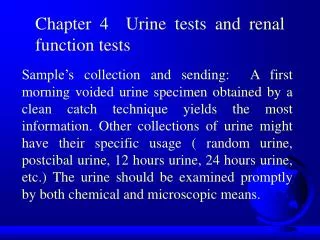
Chapter 4Renal Function Dr Atef A Masad Renal Function
What do the kidneys do? • Urine formation • Regulate body fluid osmolality and volume • Regulate electrolyte balance • Regulate acid-base balance • Excrete waste products and foreign substances • Produce and excrete hormones such as Erythropoietin and Rennin Renal Function
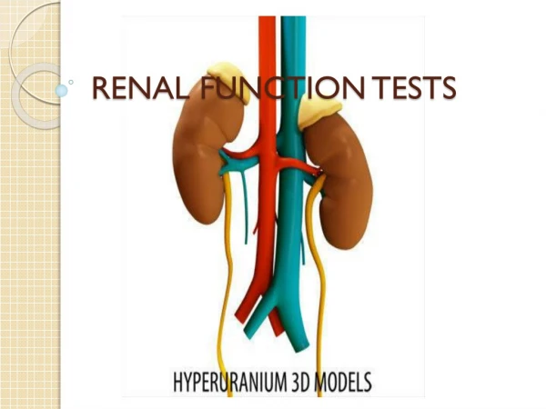
The kidneys are a pair of fist-sized organs that are located on either side of the spinal column just behind the lower abdomen (L1-3). • The kidney is a component of the urinary tract system, which consists of kidneys, ureters, urinary bladder, and urethra. • The urinary tract functions as a pathway for the elimination of metabolic by-products and unessential chemicals and removal of potentially toxic waste products. Renal Function
Renal anatomy Cortex Pelvis Capsule Medulla To the bladder Renal Function
The kidney maintains the water, ionic, and chemical balance of blood by filtering chemicals from the blood and conserving, or reabsorbing, those chemicals that are needed for adequate metabolism. • The kidneys maintain the balance of plasma constituents, while excreting those substances that are no longer needed by the body. Renal Function
The central portion of the kidney consists of tubules that drain the kidney cortex and medulla. • The cortex, or outer portion of the kidney, appears red and contains the blood vessels, which bring blood to the kidney, and nephrons, the functional units that filter and maintain the chemical stasis of the blood. • The medulla appears as a series of pyramids within the cortex and contains straight tubules and collecting ducts. Renal Function
There are about 1 million nephons in each kidney. • In association with blood vessels that serve the kidney, the nephrons make up the cortex and medulla of the kidney. • Each nephron contains a glomerulus, proximal tubule, loop of Henle, distal tubule and collecting duct. • The glomerulus filters blood plasma from arterioles into Bowman’s space and hence in the proximal tubule of the nephron. Renal Function
Proximal tubule Afferent arteriole Distal tubule Glomerulus Bowman’s capsule Collecting duct Renal artery Henle’s Loop The Nephron Renal Function
Renal Physiology • 3 basic renal processes • Glomerular filtration, • Tubular reabsorption, • Tubular secretion. • Glomerular filtration • Glomerulus filters incoming blood, all substances except cells and large molecules pass into further sections of the nephron. • Filtration process requires adequate pressure. • Water, electrolytes, glucose, amino acids, urea, creatinine pass freely and enter the proximal tubule. Renal Function
The integrity of the glomerulus membrane, which consists of the endothelium, basement membrane, and epithelium, and renal blood flow determine the glomerular filtration rate. • The glomerulus has multiple small pores through which chemicals are filtered from the blood. • In a healthy kidney, the pores exclude any substance with a molecular radius more than 4 nm. • The glomerulus also selects by charge. • Substances that are neutral or have positive charge are more likely to pass through the pores of the glomerulus than substances that are negatively charged. Renal Function
For example, albumin, which has a molecular radius of less than 4 nm but is negatively charged, does not readily pass through the pores of the glomerulus. • In a healthy kidney, cellular blood components should be excluded from the filtrate because of their size. • Albumin, many plasma proteins, cellular elements, protein-bound substances such as lipids and bilirubin are stopped. Renal Function
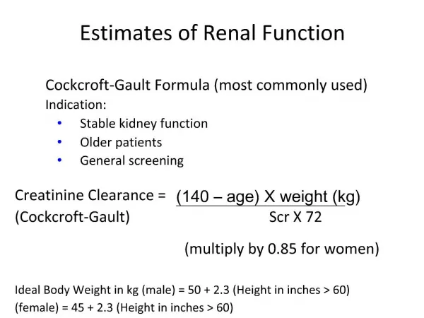
"GFR" • The kidneys receive each minute 1500-2000 ml of blood, the glomerulus filters out 125-130 ml protein and cell free. • The volume of blood filtered per minute is known as the glomerulus filtration rate Renal Function
Mean capillary blood pressure = 50 mm Hg BC pressure = 10 mm Hg Onc. pressure = 30 mm Hg Net hydrostatic = 10 mm Hg Glomerular filtration Glomerlular capillary membrane Vascular space Bowman’s space 2,000 Liters per day (25% of cardiac output) 200 Liters per day GFR 130 mL/min Renal Function
Then what happens? • If 200 liters of filtrate enter the nephrons each day, but only 1-2 liters of urine result, then obviously most of the filtrate (99+ %) is reabsorbed. • Reabsorption can be active or passive, and occurs in virtually all segments of the nephron. Renal Function
Reabsorption from glomerular filtrate Renal Function
What gets filtered in the glomerulus? • Some filtered • 2-microglobulin • 1-microglobulin • Albumin • None filtered • Immunoglobulins • Ferritin • Cells • Freely filtered • H2O • Na+, K+, Cl-, HCO3-, Ca++, Mg+, PO4, etc. • Glucose • Urea • Creatinine • Insulin Renal Function
Proximal Convoluted Tubule • It returns valuable substances back to the blood circulation, this includes ¾ of the water. • Renal threshold for each substance determines whether it is reabsorbed or secreted. • However, some substances have no renal threshold e.g H2O. • Proximal tubule secrets products of kidney tubular cell metabolism such as H+ Renal Function
Reabsorption may be active or passive • Active — against a concentration gradient (glucose, amino acids, low mw proteins, sodium, etc.) — • regulated by the kidney according to the level of these substances in the blood • Passive — no energy involved — such as water and urea • Tubular secretion may also be passive or active
Henle's Loop • The hyperosmolality (solute concentration ) is maintained by the Henle's loop. • The descending limb is permeable to water but not to solute. • Passive reabsorption of water in descending loop • The ascending limb is impermeable to water but permeable to Na, Cl and partially permeable to urea. • The medullary interstitial fluid becomes hyperosmotic compared to the fluid in the ascending limb. • The high osmolality of the surrounding interstitial medulla fluid is the physical force that accelerates the absorption of water from the descending limb. Renal Function
Na+ 300 mOsm/Kg H2O Increasing osmolality Na+ Na+ 1200 mOsm/Kg The Loop of Henle Proximal tubule Distal tubule Renal Cortex Descending loop Ascending loop Renal Medulla Renal Function
The interstitial hyperosmolality is maintained because the ascending limb continues to pump Cl– and Na+ ions into it. • The net result is production of hypoosmolal urine as it leaves the loop. • This process is called countercurrent multiplier system. Renal Function
Distal Convoluted Tubule • Small adjustments occur to achieve electrolyte and acid-base homeostasis. • It is under the control of aldosterone. • Aldosterone stimulates Na+ reabsorption by distal tubule and K and H+ ion secretion. • H+ ions secretion is linked to bicarbonate regeneration and ammonia secretion which occur here, small amounts of Cl– are reabsorbed. Renal Function
Collecting Duct • Is the final site for either concentrating urine or diluting it. • The upper portion is under the control of aldosterone which acts to stimulate Na reabsorption. • Cl and urea are absorbed here. • The collecting duct is under the control of ADH which stimulates water reabsorption. Renal Function
Water Balance • Water loss is under the control of ADH. • ADH responds primarily to changes in osmolality and intravascular volume. • Increased osmolality stimulates ADH secretion which increases the permeability of collecting tubules to water resulting in more concentrated urine. • In dehydration, reabsorption of water is increased, • In states of water excess, tubules reabsorb water at only a minimal rate resulting in excretion of large volume of dilute urine. Renal Function
Plasma hyperosmolality H2O H2O ADH (vasopressin) Regulation of H2O reabsorption Pituitary Renal Medulla (osmolality 1200 mOsm/Kg) Renal Function
Acid-Base Balance • The renal system participates in controlling body pH in addition to respiratory system and the acid-base buffering system. • The kidneys role in controlling body pH is accomplished by preserving HCO3– and removing metabolic acids. Renal Function
Regeneration of HCO3– • HCO3– are filtered by the glomerulus. • HCO3– combines with H+ in the lumen of renal tubules to form H2CO3. • H2CO3 is degraded to CO2 + H2O. • CO2 diffuses into proximal tubules and is converted to H2CO3 by the action of carbonic anhydrase, then it is degraded back to H+ and HCO3. • This regenerated HCO3 is transported into the blood to replace the depleted one by metabolism, H+ are secreted into the tubular lumen and enter the urine. Renal Function
Reaction with NH3 • NH3 is formed in the renal tubules as a result of glutamine deamination by glutaminase, NH3 then react with H+ to form NH4 which is excreted in urine. • Glutaminase • Glutamine glutamic acid + NH3 • NH3 + H+ + NaCl NH4 Cl + Na+ • This mode of acid excretion is the primary means by which the kidneys compensate for states of metabolic acidosis. Renal Function
Reaction with Monohydrogen phosphate "HPO42– • Phosphate ions filtered by glomerulus exist in tubular fluid as Na2HPo4 which can react with H+ to yield NaH2 Po4 + Na. • Na2HPo4 is excreted, it is responsible for the titratable acidity of the urine. • The released Na combines with HCO3– to yield NaHCO3 which is reabsorbed. Renal Function
Sodium is exchanged in the presence of the hormone aldosterone and water is exchanged in the presence of antidiuretic hormone (ADH). • The exchange of chemicals back into the blood supply is called reabsorption. • Exchange may occur as active transport, or as passive transport, which occurs with the gradient from high to low concentration of the chemical. • Some tubule cells, especially those in the distal portion of the nephron, exchange sodium and water back into the blood supply. Renal Function
Because of the ability of the nephron to filter and reabsorb certain chemicals from the blood, the measurement of the concentration of these chemicals in the blood and urine serves as a functional evaluation of the kidney and specific areas of the nephron. • The measurements of the concentrations of creatinine, blood urea nitrogen, and electrolytes all serve as functional evaluations of different areas of the kidney. Renal Function
Renal Endocrine Function • (A) Primary endocrine function • The kidneys synthesize rennin, prostaglandin and erythropoietin. • 1- Rennin • Rennin is produced by renal medulla whenever extracellular fluid volume decreases. • It is responsive to changes in Na+ and K+ levels in blood. Renal Function
It is vasoconstrictor which increases blood pressure. • It catalyzes the synthesis of angiotensin by means of cleavage of the circulating plasma precursor angiotensinogen.
Angiotensinogen Na+ Renin BP Angiotensin I Angiotensin II vasoconstriction Angiotensin III Adrenal cortex Aldosterone Na+ Na+ Regulation of distal tubule Na+ permeability Renal Function
ANP acts to reduce the water, sodium and adipose loads on the circulatory system, thereby reducing blood pressure, ACEangiotensin converting enzyme Renal Function
Atrial natriuretic peptide (ANP), is a powerful vasodilator hormone secreted by heart muscle cells. ANP acts to reduce the water, sodium and adipose loads on the circulatory system, thereby reducing blood pressure Renal Function
2- Prostaglandins • Prostaglandins produced by kidneys increase renal blood flow, Na and H2O excretion and rennin discharge. • They resist renal vasoconstriction due to angiotensin and norepinephrine. • Prostaglandins are used in antihypertensive therapy.
3- Erythropoietin • It is a single chain polypeptide. • It is produced by cells close to the proximal tubules. • Its production is regulated by blood oxygen levels "hypoxia increases its production". • Erythropoietin acts on the erythroid progenitor cells in the bone marrow, causing their maturation and increasing the number of RBCs. • In chronic renal insufficiency, erythropoietin production is significantly reduced causing anemia. Renal Function
(B) Secondary Endocrine Function • The kidneys are the target locus for the action of aldosterone, the catabolism of insulin, glucagons and aldosterone and as a point of activation of vitamin D metabolism. Renal Function
Renal Disorders Acute Glomerulonephritis Nephrotic Syndrome Tubular Diseases Urinary Tract Infection Acute Renal Failure 41 M. Zaharna Clin. Chem. 2009
Acute Glomerulonephritis Red cell cast Acute inflammation of the glomeruli Results in oliguria, hematuria, increased BUN and serum creatinine, decreased GFRand hypertension Red cell cast finding are of great importance Proteinuria also present 42 M. Zaharna Clin. Chem. 2009
Nephrotic Syndrome Massive proteinuria, edema, hypoalbuminemia, hyperlipidemia, and lipiduria Has many cuases Characterized by increased glomerular membrane permeability — loss of protein (greater than 2-3 grams per day) 43 M. Zaharna Clin. Chem. 2009
Tubular Diseases Depressed secretion or reabsorption of specific biochemicals Or Impairment of urine dilution and concentration mechanisms Renal Tubular Acidosis — most important Low values of phosphorus in serum, and presence of glucose and AA in urine 44 M. Zaharna Clin. Chem. 2009
Urinary Tract Infection Bladder — cystitis Kidneys — pyelonephritis Bacterial concentrations >100,000 colonies/mL Increased number of white blood cells Increased number of red blood cells may be present White blood cell casts is considered diagnostic of pyelonephritis 45 M. Zaharna Clin. Chem. 2009
Acute Renal Failure • Ocurring when the GFR is reduced to less than 10 mL/minute. • Prerenal — before blood reaches the kidney • Hypovolemia • Cardiovascular failure • Renal — occuring in kidney • Acute tubular necrosis • Glomerulonephritis • Postrenal — after urine leaves kidney • Obstruction 46 M. Zaharna Clin. Chem. 2009
Usually accompanied by oliguria Associated with varying degrees of proteinuria, hematuria, and presence of red cell casts and other casts BUN and creatinine increase rapidly Can progress to chronic renal insufficiency or failure 47 M. Zaharna Clin. Chem. 2009
Renal Calculi • Formed by the combination of various crystallized substances. • Such as Ca-phospahte, Uric Acid, Cystine, Mg-ammonium phosphate and Ca-oxalate • Calcium oxalate stones are the most common. • Formed due a reduced urine flow rate due to decrease fluid intake and urine saturation of insoluble substances, ifections, Gout, inherited diseases, Hyperparathyroidism, high urine Ca, Vitamin D toxicity
Chemical analysis is available and x ray diffraction. • Clinical symptoms: hematuria, UTI, and abdominal pain