
Target & Strategy
(a). (c). (b). (b) 0.16 µm. (a) PMMA spin-casted film. (c) 0.65 µm. (d). (e). (a). (b). (d) 0.97 µm. (e) 8.64 µm. (c). (d). Relative modulus of different substrates: PMMA spin-casted film, aligned fibers and random fibers.

Target & Strategy
E N D
Presentation Transcript
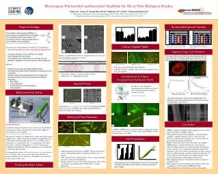
(a) (c) (b) (b) 0.16 µm (a) PMMA spin-casted film (c) 0.65µm (d) (e) (a) (b) (d) 0.97µm (e) 8.64µm (c) (d) Relative modulus of different substrates: PMMA spin-casted film, aligned fibers and random fibers Relative modulus of AHDFs on the different substrates: PMMA spin-casted film aligned fibers and random fibers Optical Image SEM Image Electrospun Poly(methyl methacrylate) Scaffolds for 3D in-Vitro Biological Studies Ying Liu1, Yuan Ji1, Kaustabh Ghosh2, Richard A.F. Clark2, Miriam Rafailovich1 1. Department of Materials Science and Engineering, SUNY at Stony Brook, Stony Brook, NY 11794-2275 2. Department of Biomedical Engineering, SUNY at Stony Brook, Stony Brook, NY 11794-8181 Target & Strategy Surface Mechanical Property Poly (methyl methacrylate) (PMMA) is a rigid polymer biomaterial with good degree of compatibility with human tissue. It is commonly used for making contact lenses, bone implants, and prosthetics. (Li SH, De Wijin JR, et al. Journal of biomedical materials research 61(1):109-120 2002) Cell on Aligned Fibers • Can one use this polymer to create a 3D scaffold to direct cell growth for tissue engineering application? • The large modulus of the scaffold can support physiologically relevant loads • The scaffold can be patterned for orienting the cell adhesion, migration and extra-cellular matrix deposition Agarose Drop Cell Migration Radius is controlled by the solvent, vapor pressure, viscosity, and electric field lines (dielectric constant). (Dan Li, Younan Xia.Adv.Mater. 2004, 16, No.14) • Cell aspect ratio and cell density were measured after 24 hours incubation and plotted against the distance from the edge of the agarose droplet. Bars, 0.5 mm (a), 100 mm (b). • Method • Prepare non-woven 3D febrile scaffold using electrospinning where fiber diameter and spacing can be controlled. • Culture human dermal fibroblasts and compare two dimensional films and three dimensional scaffolds regarding: • Orientation • Cell proliferation • Focal adhesion points expression • Cell migration PMMA spincasted film PMMA random mesh PMMA alinged mesh • Cells were oriented along the fiber direction. • If the fiber density was high, some cells were extended to span two adjacent fibers. • Electrospun “ribbons” resulted in all the solvents • Aspect ratio ~3, independent of radius Cell Adhesion to Fibers: Imaging Focal Adhesion Points Aligned Fibers In order to assess the spatial distribution of focal adhesion contacts, cells were fluorescently stained to visualizevinculin. Molecular biology of the cell, 4th edition Electrospinning Setup • (a) On the single fiber, and (b) cross-aligned fiber, the filopodia of the cells could sense the environment around the cells. Bars, 20 mm (a, b). We produced a cross-aligned 3D scaffolds by spinning the PMMA fibers at a rate of 700 RPM in two mutually perpendicular directions on the same substrate. Effects of Fiber Diameter Conclusion Electrospinning: a method of using electrostatic forces to form very fine filaments from polymer solution. Its main advantage include simplicity, cost-efficiency, and scalability. (Y.Z.Zhang, J, Venugopal et al. Biomacromolecules 2005, 6, 2583-2589) The polymer solution was loaded in the syringe and 5-8kV voltage supplied by (a) a high-voltage power supply was applied between (b) the needle and the ground. The flow rate of solution (about 20mL/min) was controlled by the (c) syringe pump. Two targets were used, a stationary aluminum disc or (d) a rotating drum wrapped in aluminum foil. The fiber alignment was controlled by (e) the motor speed rotation. By changing the polymer solution ( the solvent, the concentration et al.), as well as the rotating speed, the average diameter of polymer filament can be varied in a controllable way, from nano-meter to micro-meter scale. (a) AHDFs on PMMA spin-casted film; random scaffolds with diameter of (b) 0.16µm, (c) 0.65µm, (d) 8.64µm and (e) aligned PMMA scaffold with fiber diameter of 8.64µm • PMMA scaffolds with different fiber diameters can be electro-spun from CHCl3 and DMF solutions. • Fiber diameter affected cell morphology: 160 nm diameter scaffolds resulted in dentritic cells as well as flat cells. However, when the fiber diameter above 1 micron, the cell grow along the direction of the fiber. • Highly oriented cells were observed on the cross-aligned PMMA fibrous scaffold with diameter of 8.64 micron. • The focal adhesion points for cell on the fiber with diameter above 1 micron, were concentrated along the oriented fiber diameters. Cells on the fibers with diameter less than 1 micron, had focal adhesion points uniformly distributed along the cell body. On the control substrates focal adhesion points were concentrated along the cell periphery and nucleus. • Fiber alignment increase cell proliferation due to improved packing of cells. • Fiber alignment does not influences the stiffness of the cells comparing to cells growing on PMMA thin film • Fiber aligned increase the direction of the cell migration. Cell Proliferation • Adult human dermal fibroblast (AHDF) cell body followed the course of the underlying fibers with fiber diameter above 1 micron . • Cells cultured on the 160nm diameters fibers assumed dendritic structure as well as flat morphology. • Similar features were also observed by Grinnell for cells seeded on collagen extracellular matrix (ECM) fibers of similar dimensions. • (Frederich Grinnell, Chin-Han Ho et al. Molecular biology of the cell, Vol.14,384-395,2003) • Cells packing is more efficient and hence do not become confluent as quickly. Hence effect is more pronounced after 9 days. Finding the Right Fibers