Download
1 / 47
480 likes | 863 Views
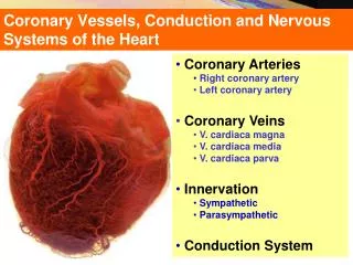
Coronary Vessels, Conduction and Nervous Systems of the Heart. Coronary Arteries Right coronary artery Left coronary artery Coronary Veins V. cardiaca magna V. cardiaca media V. cardiaca parva Innervation Sympathetic Parasympathetic Conduction System.

E N D
Coronary Vessels, Conduction and Nervous Systems of the Heart • Coronary Arteries • Right coronary artery • Left coronary artery • Coronary Veins • V. cardiaca magna • V. cardiaca media • V. cardiaca parva • Innervation • Sympathetic • Parasympathetic • Conduction System
CoronaryArteries-Arteries of the Heart Coronary arteries: Supply blood to the myocardium and epicardium. Start from the right and left aortic sinuses. Anastomoses between the branches of the coronary arteries enables the development of the collateral circulation.
CoronaryArteries Right coronary artery: Runs in the coronary groove. Gives off conal branches. Gives of the SA nodal (may arise from the circumflex branch of left) and the AV nodal branches as well as the right marginal branch and posterior interventricular artery (runs in the posterior interventricular groove). Terminates by anastomosing with the circumflex branch of the left coronary artery.
CoronaryArteries Right coronaryartery: Originates above the right cusp of the aortic valve. Travels down the right atrioventricular groove, towards the crux of the heart. Branches into the posterior descending artery and the right marginal artery. At the origin of the RCA is the conus artery. In addition to supplying blood to the right ventricle (RV), the RCA supplies 25% to 35% of the left ventricle (LV).
CoronaryArteries Right coronaryartery: In 85% of patients (Right Dominant), the RCA gives off the posterior descending artery (PDA). In the other 15% of cases (Left Dominant), the PDA is given off by the left circumflex artery. The PDA supplies the inferior wall, ventricular septum, and the posteromedial papillary muscle. The RCA also supplies the SA nodal artery in 60% of patients. The other 40% of the time, the SA nodal artery is supplied by the left circumflex artery.
CoronaryArteries Left coronary artery: Runs in the coronary groove. Terminates by dividing into its two branches: The anterior interventricular artery Diagonal artery The circumflex branch The left marginal artery (posterior left ventricular branch)
CoronaryArteries Left coronary artery: Arises from the aorta above the left cusp of the aortic valve and feeds blood to the left side of the heart. Abbreviated asLCA and also known as the left main coronary artery (often abbreviated LMCA) Typically runs for 1 to 25 mm and then bifurcates into the anterior interventricular artery (also called left anterior descending (LAD)) and the left circumflex artery (LCX).
CoronaryArteries Left coronary artery: Sometimes an additional artery arises at the bifurcation of the left main artery, forming a trifurcation; this extra artery is called the intermediate artery. The part that is between the aorta and the bifurcation only is known as the left main artery (LM), while the term 'LCA' might refer to just the left main, or to the left main and all its eventual branches.
CoronaryArteries LCA: Left Coronary Artery, RCA: Right Coronary Artery (=MARG: Marginal branch), CB: Circumflex branch, LAD: Left Anterior Descending (=AIB:Anterior interventricular branch), DIAG: Diagonal interventricular branch of anterior interventricular artery, RCA: Right coronary artery, AB: (Right) Atrial branch, SANB: Sinuatrial nodal branch, RMA: Right marginal branch, LMA: Left marginal branch, ACV: Anterior cardiac vein SCV: Small cardiac vein, GCV: Great cardiac vein (=AIV: Anterior interventricular vein), MCV: Middle cardiac vein, SCV: Small cardiac vein, PIA: Posterior interventricular branch (=PDA: Posterior descending artery), AVN: Atrioventricular nodal (branch), CS: Coronary sinus, IVC: Inferior venacava
CoronaryArteryDominance • The coronary artery that supplies the posterior interventricularartery determines the coronary dominance. • If it is supplied by the right coronary artery, then the coronary circulation can be classified as "right-dominant". • If it is supplied by the circumflex artery (CX), a branch of the left artery, then the coronary circulation can be classified as "left-dominant". • If it is supplied by both the right coronary artery (RCA) and the circumflex artery, then the coronary circulation can be classified as"co-dominant". • Approximately 70% of the general population are right-dominant.
Coronary(Cardiac) Veins Great cardiac vein (anterior interventricular vein) Middle cardiac vein (posterior interventricular vein) Small cardiac vein (right side) Oblique vein of the left atrium Anterior cardiac veins Smallest cardiac veins
Coronary(Cardiac) Veins • Greatcardiacvein(anteriorinterventricularvein = v. cordis magna) • The Great Cardiac Vein (left coronary vein) begins at the apex of the heart and ascends along the anterior longitudinal sulcus to the base of the ventricles. • It then curves to the left in the coronary sulcus, and reaching the back of the heart, opens into the left extremity of the coronary sinus. • It receives tributaries from the left atrium and from both ventricles: one, the left marginal vein, is of considerable size, and ascends along the left margin of the heart.
Coronary(Cardiac) Veins • Middlecardiacvein(posteriorinterventricularvein= v. cordis media) • The middle cardiac vein commences at the apex of the heart. • Ascends in the posterior longitudinal sulcus/ • Ends in the coronary sinus near its right extremity. • .
Coronary(Cardiac) Veins • Smallest cardiac veins(Thebesian veins= veins of Thebesius) • Thebesian veins are minute valveless veins in the walls of all four heart chambers. • They are most abundant in the right atriumand least in the left ventricle. They drain the myocardiumand pass through the endocardial layer to empty mostly into the right atrium, but a few empty into the ventricles. • The openings of the chambers are called the foramina venarum minimarum. • The Thebesian venous network is considered an alternative (secondary) pathway of venous drainage of the myocardium. • They are named after the German anatomist Adam Christian Thebesius,who described them in a 1708 treatise called Disputatio medica inauguralis de circulo sanguinis in corde.
Coronary(Cardiac) Veins • Smallcardiacvein(right coronary vein = v. cordisparva = v. cardiacaparva) • The small cardiac vein runs in the coronary sulcusbetween the right atrium and ventricle. • Opens into the right extremity of the coronary sinus. • Receives blood from the back of the right atrium and ventricle. • It may drain to the coronary sinus, right atrium, middle cardiac vein, or be absent.
Lymphatic Drainage of the Heart • Tracheobronchial lymph nodes: situated above or below the bifurcation of trachea.
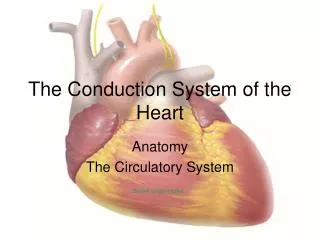
ConductionSystemof theHeart Composed of specializedcardiacmusclefibersthatproduceimpulsesandconductthemthroughoutthemyocardium. SA (sinuatrial) node Localized in thesuperiorpart of the terminal crest. Pacemaker of theheart Gives of impulsesabout 70 timesperminute. Impulsesfrom SA nodespreadsthroughthemuscularwall of theatriaandreachesthe AV node. AV (atrioventricular) node Located on the ‘interatrialseptum’ slightlysuperiortotheopening of thecoronarysinus Transmittheincomingsignalstothe AV bundle AV bundle Is theonlybridgebetweentheatrialandventricularmyocardium Dividesintorightandleftbundles.
ConductionSystemof theHeart SA (sinuatrial) node Localized in the superior part of the terminal crest. Pace maker of the heart Gives of impulses about 70 times per minute. Impulses from SA node spreads through the muscular wall of the atria and reaches the AV node.
ConductionSystemof theHeart SA (sinuatrial) node Localized in the superior part of the terminal crest. Pace maker of the heart Gives of impulses about 70 times per minute. Impulses from SA node spreads through the muscular wall of the atria and reaches the AV node.
ConductionSystemof theHeart AV (atrioventricular) node Located on the ‘interatrialseptum’ slightly.superiortotheopening of thecoronarysinus Transmittheincomingsignalstothe AV bundle.
ConductionSystemof theHeart Composed of specializedcardiacmusclefibersthatproduceimpulsesandconductthemthroughoutthemyocardium. SA (sinuatrial) node Localized in thesuperiorpart of the terminal crest. Pacemaker of theheart Gives of impulsesabout 70 timesperminute. Impulsesfrom SA nodespreadsthroughthemuscularwall of theatriaandreachesthe AV node. AV (atrioventricular) node Located on the ‘interatrialseptum’ slightlysuperiortotheopening of thecoronarysinus Transmittheincomingsignalstothe AV bundle AV bundle Is theonlybridgebetweentheatrialandventricularmyocardium Dividesintorightandleftbundles.
Conduction System of the Heart 2 1 4 5 3
Innervation of the Heart The medulla receives sensory input from different systemic and central receptors (e.g., baroreceptors and chemoreceptors). Autonomic outflow from the brainstem is divided principally into sympathetic and parasympathetic (vagal) branches. Efferent fibers of these autonomic nerves travel to the heart and blood vessels where they modulate activity of these target organs. The S-A node is innervated by vagal (parasympathetic) and sympathetic fibers. Sympathetic efferent nerves are present throughout the atria (especially in the S-A node) and ventricles, and in the conduction system of the heart.
Innervation of the Heart The medulla receives sensory input from different systemic and central receptors (e.g., baroreceptors and chemoreceptors). Autonomic outflow from the brainstem is divided principally into sympathetic and parasympathetic (vagal) branches. Efferent fibers of these autonomic nerves travel to the heart and blood vessels where they modulate activity of these target organs. The S-A node is innervated by vagal (parasympathetic) and sympathetic fibers. Sympathetic efferent nerves are present throughout the atria (especially in the S-A node) and ventricles, and in the conduction system of the heart.
Innervation of the Heart Innervation of the heart is through the autonomic nerves (both sympathetic and parasympathetics) from the cardiac plexus. Cardiac plexus lies posterior to the ascending aorta and bifurcation of the pulmonary trunk. Sympathetics come from the cervical and superior 5 thoracic sympathetic ganglia. Parasympathetics come from the vagus nerves.
Cardiac Referred Pain Ischemia of the myocardium produces cardiac pain (referred as angina or angina pectoris). Axons of the GVA fibers enter the spinal cord through T1 - T5 segments (mostly on the left side). Cardiac referred pain is a phenomenon in which the noxious stimuli originating in the heart are felt as pain arising from the related dermatomes.
Innervation of the Heart • Parasympathetic effect • Decreases heart rate. • Diminishes blood pumping force of the heart. • Constricts coronary arteries • Sympathetic effect • Increases heart rate. • Strengthens blood pumping force of the heart. • Dilates coronary arteries.
Angina Pectoris • Over eating • Exhausting exercise • Cold • Smoking
Projection of the Heart Valves on the Chest Wall 1 2 3 Pulmonary valve Aortic valve 4 5 Mitral valve Tricuspid valve 6
Ouscultation of the Heart 1 2 Pulmonary valve (Left-2.ICS) 3 Valva aortae (Right- 2. ICS) 4 5 Tricuspid valve (Right-6.CC) Valva mitralis (Left-5.ICS-MCL) 6