Bone Classification
Bone Classification(cont'd). Long bones: length exceeds width;shaft

Bone Classification
E N D
Presentation Transcript
1. Bone Classification 206 named bones
Axial skeleton
Appendicular skeleton
Shape classification: long, short, flat, irregular, sesamoid
2. Bone Classification(cont�d) Long bones: length exceeds width;shaft & 2 ends;primarily compact w/spongy interior; ex. humerus, femur
Short bones: cubelike;spongy bone; ex. carpals, tarsals
Flat bones: thin,flattened, w/slight curvature;compact bone surfaces w/spongy layer; ex. sternum, ribs
3. Bone Classification(cont�d) Irregular bone: complicated shapes & mostly spongy bone; ex. vertebra, pelvis
Sesamoid:short bone,forms within tendon;patella
4. Bone Functions Support-hard framework;supports body wall (limbs, rib cage)
Protection-braincase, vert.foramina
Movement-levers
Storage
Blood cell formation
5. Bone Structure Bones are organs-osseous tissue, along with nervous,cartilaginous,fibrous CT
Osteocytes, osteoblasts, osteoclasts
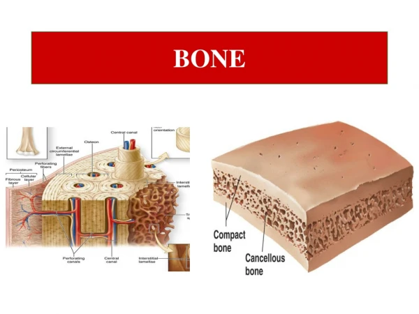
6. Textures: Compact vs Spongy
Compact-dense, smooth,solid outer layer
Spongy bone-honeycomblike; trabeculae
7. Structure of Typical Long Bone Diaphysis-compact bone surrounds cavity;yellow marrow evident in adults
Epiphyses-compact exterior,spongy interior;hyaline cartilage on joint surface
8. Structure of Typical Long Bone (cont�d) Periosteum-double layered (outer & inner);fibrous outer, inner has osteoblasts & osteoclasts;Sharpey�s fibers
Endosteum-lines marrow; osteoblasts & osteoclasts
9. Structure of short, irregular & flat bones Non-cylindrical
No marrow cavity
Dipl�e-internal layer of spongy bone in flat bones
10. Hematopoietic Tissue Red marrow
In newborns, red marrow predominate cavities
Adults: RBC produced in femoral& humeral head, diploe of sternum, & irregular bones (pelvic)
11. Microscopic Structure of Bone Compact bone-has osteons
Osteon-has Haversian system
Haversion system-central canal, Volkmann�s canal, lacunar osteocytes, & canaliculi
Spongy bone
12. Chemical Composition of Bone Organic components-Osteoblasts, osteocytes, osteoclasts;glycoproteins & collagen fibers
Inorganic components-hydroxyapatites (Ca phosphate/hydroxide),Ca carbonate & ions
Organic/inorganic combo gives durability/strength w/o being brittle
13. Bone Markings Muscle & ligament attachment projections-tuberosity, crest, line, tubercle, trochanter, epicondyle, spine
Joint forming projections-head,facet, condyle, ramus
Depressions/openings for blood vessels & nerves-meatus, groove, fossa, foramen
14. 80 bones
The Skull
Vertebral Column
Bony Thorax
15. Neurocranium (8)-Enclose brain and protect organs of hearing and equilibrium.
Viscerocranium (14)- (1) Forms facial framework;(2) Provide cavities for the sense organs of sight, taste, and smell;(3) Provide openings for passage of air and food;(4) Secure the teeth; (5) Anchor facial muscles of expression.
16. 1 Frontal bone -anterior portion of cranium;the forehead and roofs of the orbits.
Orbits
Anterior cranial fossa
Glabella
Frontal sinuses
17. 2 Parietal Bones- Large,curved, rectangular bones forming superior and lateral aspects of the skull; largest sutures occur at parietal bone articulation points.
Major Sutures-(1) Coronal suture- parietal bones meet with frontal bone anteriorly. (2) Sagittal suture-right and left parietals meet superiorly at cranial midline.
18. (3)Lambdoid suture-the parietal bones meet the occipital bone posteriorly.
(4)Squamous suture-parietal and temporal bone meet on lateral aspect of skull.
19. 1 Occipital Bone -posterior wall and base of the skull
internally forms walls of posterior cranial fossa
foramen magnum
occipital condyles
external occipital protuberance (�occiput�).
20. 2 Temporal Bones -inferolateral aspects of the skull and partial cranial floor; four regions are squamous, tympanic, mastoid, petrous; zygomatic process and arch, mandibular fossa,external acoustic meatus, styloid process, mastoid process, middle cranial fossa, middle/inner ear cavities.
21. 1 Sphenoid Bone -Keystone of cranium that forms central wedge; greater/lesser wings, pterygoid processes.
Sella turcica (hypophyseal fossa)
Optic canals, superior orbital fissure
Orbital wall (lateral)
22. 1 Ethmoid -Complex shaped, lies between sphenoid and nasal bones,most deeply situated bone of the skull
Cribiform plate
Crista galli (dura mater attachment)
Perpendicular plate
Superior/middle nasal conchae
Orbital wall (medial)
23. 1 Mandible -Largest,strongest, facial bone.
Body-forms the chin
Rami-meet with body posteriorly to form angle.
Mandibular notch separates coronoid process & mandibular condyle.
Mandibular, mental foramina
24. 2 Maxillary bones- Keystone bones of the face;form upper jaw & central portion of facial skeleton.
Incisive foramen
Infraorbital foramen
Maxillary sinuses-Largest of paranasal sinuses
25. 2 Zygomatic Bones � �Cheekbones�;articulates with temporal bones via zygomatic arch.
2 Nasal Bones-Thin rectangular bones fused medially; forms �nosebridge�; inferiorly attach to nasal cartilages.
26. 2 Lacrimal Bones -Delicate fingernail-shaped bones that contribute to the medial walls of each orbit; lacrimal fossa houses lacrimal sac.
2 Palatine Bones-Forms posterior part of the hard palate.
27. 1 Vomer -Slender,plow shaped bone that lies in the nasal cavity and forms part of the nasal septum.
2 Inferior nasal conchae -Thin, curved bones of nasal cavity; inferior to middle nasal concha of ethmoid; largest of the three pairs of conchae.
28. Orbits formed by tributary bones:Frontal, Sphenoid, Ethmoid, Zygomatic, Maxillary, Lacrimal, and Palatine (Fig.7.9)
Nasal Cavity-Roof formed by cribiform plate; lateral walls formed by nasal conchae, floor formed by palatine process of maxillary bone and palatine bones.
Paranasal cavities-frontal,sphenoid,ethmoid, maxillary.
29. Does not articulate directly with any other bone in the body.
Greater horn supports larynx, acts as movable base for tongue.
Lesser horn are attachments for stylohyoid ligaments
30. Comprised of 26 irregular bones
Axial support of the trunk
Spinal cord surrounded by vertebral
foramen
Provides attachment points for the ribs and back muscles
31. Supporting ligaments are the anterior/posterior longitudinal ligaments.
Intervertebral discs are cushionlike paddings; inner semifluid nucleus pulposus and a strong outer ring of fibrocartilage called the annulus fibrosus.
Discs accounts for 25% of vertebral height.
Herniated disc is the rupturing of the annulus fibrosus.
32. Cervical
Thoracic
Lumbar
Sacrococcygeal
33. Primary ( Thoracic & Sacral)
Secondary ( Cervical & Lumbar)
Kyphosis
Lordosis
Scoliosis
34. Body
Vertebral arch (lamina & pedicles)
Vertebral foramen
Spinous/Transverse process
Superior/Inferior articular processes/ facets
Intervertebral foramina
35. �Typical�(C3-C7) has oval body, short bifid spinous process, and transverse foramina.
Vertebra prominens
1st (atlas) (no body, no spinous process, superior articular facets �carry� the skull)
2nd one is the axis (has body, spinous process, and dens)
36. Increase in size from the first to last.
Heart shaped body,
Circular vertebral foramen.
Costal facets(on TPs)
37. Large bodies
Short laminas and pedicles
Short & flat spinous processes
Superior/inferior articular processes modified to�lock� preventing rotation of lumbar spine.
38. Formed by five fused vertebrae (in adults)
Auricular surface (sacroiliac joint)
Shapes the posterior wall of the pelvis
Two wing like alae
Sacral promontory
Transverse lines
Sacral foramina
Median & lateral sacral crests
Sacral canal & hiatus
39. Vestigial tailbone
Attachment site for ligaments and sphincter muscle
Four or five fused vertebrae (completed in late adulthood)
Gender positions
40. Forms protective cage around vital organs of the thoracic cavity (heart, lungs, and great blood vessels).
Supports the shoulder girdles and upper limbs.
Provides attachment points for the muscles of the back, chest, and shoulders.
Intercostal spaces between the ribs are occupied by intercostal muscles.
41. Flat bone approximately 15cm.long (6 in.)
Fusion of three bones: manubrium, body, and xiphoid process.
Landmarks: jugular notch,sternal angle and xiphisternal joint.
42. Ribs originate on/between thoracic vertebrae; attach to sternum
12 pairs
7 true (vertebrosternal)
3 false (vertebrochondral)
2 floating(vertebromuscular ribs)
Rib morphology: head, neck, tubercle,angle, shaft, costal groove.
43. The pectoral(shoulder) girdle and upper limb
The pelvic (hip)girdle and lower limb
44. Clavicles: Direct connection between pectoral girdle/axial skeleton;slender doubly curved long bones; have acromial and sternal ends.
Scapulae: Thin, triangular flat bones; important structures are:borders (sup., med.,lat.), spine, acromion (ac joint),glenoid cavity, coracoid process, supra/infra spinous fossae,and subscapular fossa.
45. Humerus: Articulates with glenoid cavity at the scapula and with ulna/radius at the elbow; important structures are: head,surgical neck, greater/lesser tubercles;capitulum, trochlea, coronoid and olecranon fossae, lateral and medial epicondyles.
46. Ulna: Slightly longer than radius & medial; important structures are: olecranon and coronoid processes, trochlear notch, ulnar head and styloid process.
Radius: Lateral; important structures are the radial head and styloid process.
Antebrachial interosseous membrane
Pronation/supination
47. Proximal bones (medial to lateral) Scaphoid Lunate Triquetral Pisiform
48. Distal bones(medial to lateral) Trapezium
Trapezoid
Capitate
Hamate
49. Metacarpals (Palm): 5 small long bones; Roman numerals(I-V) used to identify; proximal �base�, �body�, distal �head�; heads are what make up the �knuckles�.
Phalanges (Fingers): 14 miniature long bones; pollex = thumb; all except pollex have proximal,middle, and distal phalanges.
50. Comprised of three fused bones: The ilium, ischium, and pubis
Ilium: Superior region;important structures are: iliac crest, anterior/posterior superior iliac spines, anterior/posterior inferior iliac spines.
Ischium:Posteroinferior region;ischial spine, ischial tuberosity;lesser sciatic notch.
Pubis:Superior/inferior rami, pubic symphysis, pubic arch;forms obturator foramen(isch./pubis)
51. See table 7.4
False pelvis- Portion of pelvis superior to pelvic brim.
True pelvis-Portion of pelvis inferior to pelvic brim; forms deep bowl containing the pelvic organs.
52. Femur- Largest, longest, strongest bone in the body;length is 1/4th of a person�s height; articulates with hip.Important structures are: fovea capitis, head, neck (weakest), greater/lesser trochanters,linea aspera,lateral/medial condyles, patellar surface,
Knee-patella
53. Tibia- 2nd largest, longest, strongest bone in body;important structures are: the medial/lateral condyles, intercondylar eminence (with tubercles),tibial tuberosity, anterior crest, medial malleolus.
Fibula- Sticklike bone with slightly expanded ends; the head and its lower end is the lateral malleolus.
Crural interosseous membrane
54. Talus-transmits weight of body from tibia towards toes;2nd largest foot bone.
Calcaneus-largest of tarsal bones; posterior surface attaches calcaneal tendon.
Cuboid bone
Navicular
Cuneiforms-medial, intermediate, lateral.
55. Metatarsals-1st metatarsal supports weight of body.
Phalanges-14 bones organized anatomically the same as fingers; hallux=big toe