Download
1 / 158
1.62k likes | 2.91k Views
Anathomy. The gastrointestinal tract possess a broadly similar structure throughout its length an innermost epithelium a subepithelial lamina propria

E N D
Anathomy • The gastrointestinal tract possess a broadly similar structure throughout its length • an innermost epithelium • a subepithelial lamina propria • two muscle layers, an inner circular and an outer longitudinal layer, between which lies the myenteric plexus, the intrinsic neural control system of the musculature • While this description most accurately describes the small intestine, the other organs of the gastrointestinal tract differ only subtly from this stereotype.
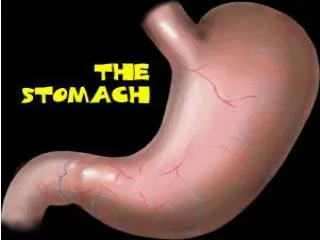
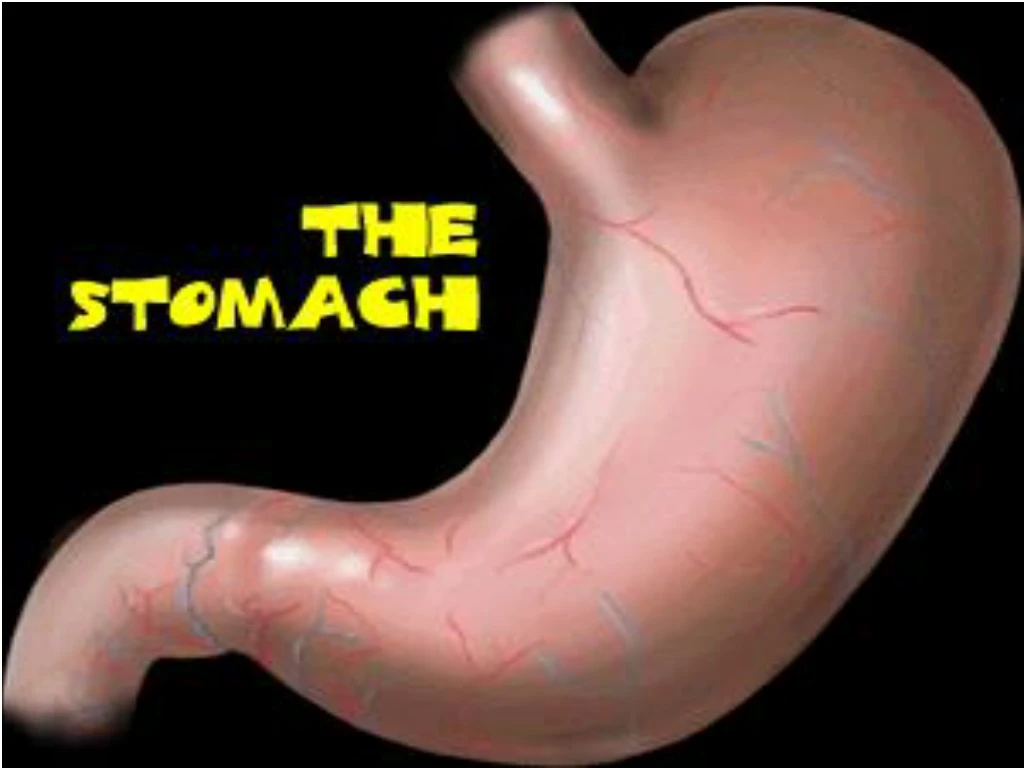
The anatomy of the stomach differs from the intestine, possessing: • an additional oblique muscular layer • at either end a sphincter—specialized musculature designed to act as a unidirectional valve to control the flow of luminal contents • the sphincter between the oesophagus and stomach (the lower oesophageal sphincter) lies at the level of the diaphragm • the sphincter between the stomach and small intestine is known as the pylorus.
Proceeding from the surface epithelium into the pits, the mucosal cells considered in detail in the following subsections include the following: • - mucous cells and • mucous neck cells, which line the surface and extend into the pits, respectively; • parietal cells, which secrete hydrochloric acid and intrinsic factor; • endocrine cells, • which secrete a variety of mediators; • - toward the base of the pits, chief cells, which secrete pepsinogens • Mucous cells.
The acid-secreting parietal cell is located in the oxyntic gland, adjacent • to ECL cell, D cell, important in the • gastric secretory process • This unique cell also secretes intrinsic factor (IF). • The parietal cell expresses receptors for several • stimulants of acid secretion including histamine (H2), gastrin (cholecystokininB/gastrin receptor), and acetylcholine (muscarinic, M3) • Each of these are G protein–linked • Binding of histamine to the H2 receptor leads to activation of adenylatecyclase and an increase in cyclic AMP. • Activation of the gastrin and muscarinic receptors results in activation of the proteinkinaseC/phosphoinositide signaling pathway. • Each of these signaling pathways in turn regulates a series of downstream kinase cascades, which control the acid-secreting pump, H, K-ATPase. • Parietal cells also express receptors for ligands that inhibit acid production (prostaglandins, • somatostatin, and EGF).
The duodenum (so named because it is 12 fingers' breadth in length) • retroperitoneal • possess on its medial aspect the ampulla of Vater which connects the pancreatic and common bile ducts to the duodenal lumen
Duodenal juice • an alkaline protein-containing fluid of weak digestive power that is secreted by the duodenum • contains invertase, maltase, lactase, erepsin, and enterokinase • the chyme passes out of the stomach with an acid reaction, and its undigested constituents are at once subjected to a second process of digestion in the duodenum by an alkaline fluid, which is a mixture of the pancreatic juice, the bile, and the enteric juice. • the pancreatic juice converts the remaining starch into sugars, and the remaining proteids into peptones, leucin, tyrosin, and fatty acids • the bile it partly emulsifies and partly saponifies the fats • the sugars are converted into lactic acid and butyric acid, possibly in part by the succusentericus, which is also amylolytic.
Defensive mechanisms • The properties of the surface epithelial cells that provide the second line of gastroduodenal defensive factors include restitution, epithelial cell metabolism (e.g., transmembrane, transcellular resistances), acid-base transporters that maintain intracellular pH, and mucus secretion. • The key subepithelial process that prevents mucosal injury is adequate mucosal blood flow.
Common symptoms • Dyspepsia • Nausea • Vomiting • Bloating • Fast enough
Bloating Indigestion Pain in Epigastric Region
Patients may speak of: • indigestion (to describe any low-grade upper abdominal discomfort) • sickness (to describe either nausea or vomiting)
Dyspepsia • upper abdominal or lower chest discomfort or pain related to eating which may be described by the patient as a burning, a heaviness, or an aching • often accompanied by other symptoms such as nausea, fullness in the upper abdomen, or belching • the symptoms of upper gastrointestinal disease are imprecise and non-specific - the clinical history will often facilitate making the correct diagnosis quickly and limit unnecessary investigation
Functional dyspepsia • previously called non-ulcer dyspepsia • dyspepsia "without evidence of an organic disease that is likely to explain the symptoms“ • is estimated to affect about 15% of the general population in western countries
Nausea • the term nausea should be restricted to the feeling of being about to vomit. • acute nausea is usually accompanied by hypersalivation. • caused by labyrinthine stimulation (as in motion sickness), distension of hollow viscera, or any severe somatic pain and by some drugs, especially opiates and those used in chemotherapy for malignant conditions
Vomiting • forceful ejection of gastric contents through the mouth by the co-ordinated contraction of abdominal and gastric muscles with relaxation of the lower oesophageal sphincter. • non-productive vomiting is called retching. • Vomiting occurs with peptic ulceration, especially when there is delayed gastric emptying (pyloric stenosis), and with advanced gastric cancer. • It occurs with disorders of the biliary tree (especially as a result of gallstones) and with acute pancreatitis (in which it is a prime symptom). • It is an important symptom of intestinal obstruction, especially with lesions above the ileocaecal valve, and it may occur with any cause of peritoneal inflammation such as appendicitis. • Metabolic causes of vomiting include diabetic ketoacidosis, hypoadrenalism, and uraemia. • Drugs which cause vomiting include opiates, some antibiotics (for example erythromycin), and chemotherapeutic agents. • Alcoholism, raised intracranial pressure, and pregnancy are important causes of early morning vomiting.
Effortless vomiting • without a definable cause may be psychogenic • this is usually a disorder of young women many of whom have suffered psychological trauma (such as sexual abuse) • it is not related to the vomiting of bulimia, a condition that is part of the anorexia nervosa syndrome • Rumination is the repetitive regurgitation of gastric contents into the mouth after meals, the regurgitated material then being reswallowed. It is not associated with nausea, heartburn, or discomfort and often appears to be simply an acquired habit.
Abdominal pain • Pain in the upper abdomen has been considered under the heading dyspepsia. Upper abdominal discomfort is so common that its presence alone is of no value in distinguishing between those patients with organic disease and those with a functional disorder. • Symptoms are rarely specific but should be reinterpreted in the light of screening investigations (such as the blood count, a straight radiograph of the abdomen, and assessment of serum markers of inflammatory disease).
Investigations • Upper GI endoscopy • Investigation for Helicobacter pylori • Find medication-related dyspepsia
A gastrointestinal ulcer is defined as a breach in the epithelium that penetrates the muscularismucosae. • If the muscularis is not breached it is called an erosion. • Duodenal ulcers and gastric ulcers are often considered together as peptic ulcers but differ considerably with regard to epidemiology, pathogenesis, presentation, and • management
Epidemiology • Developed countries: 1980-1999 • DU 8-10% and annual incidence of 0,2% • GU 3 times more rare • Decrease in incidence since 1970 • Decrease of DU principally in men M/F 3-4/1 la 2/1 and even 1/1 • GU no significant modifications in incidence. • Decreased incidence principally in young and middle aged men; is becoming a disease of old age.
Etiopathogenesis • Aggressive factors implicated in ulcer • Acid hypersecretion • Increase in parietal cell mass • Increase in vagus tone • Increase in parietal cellular sensibility to gastrin • Antral G cells hyper function • Nocturnal acid hypersecretion • Deterioration in inhibitory mechanism of acid secretions • Motility disturbances: Duodenal ulcer-rapid gastric emptying, gastric ulcer- gastric hypomotility • Pepsin hypersecretion • Hiperpepsinogenemia I - Helicobacter pylori • Duodenogastric reflux • Biliary acids, lisolecitin and proteolytic enzymes.
Etiopathogenesis • Factors of defense • Pre-epithelial factors • Bicarbonate and mucus barriers • Tensioactive phospholipids • Epithelial factors • Cellular resistance (normal cellular metabolism) • Intra-cellular PH maintenance • Growth factors (epithelial growth factors, Prostaglandins, NO) • Mechanism of repair of epithelial lesions • Post-epithelial factors • Abnormal mucous blood flow
Etiopathogenesis • Individual factors • Genetic factors: group 0 & A, Lewis and non-secretory factor (a-b-) • Familial Hyperpepsinogenemia type I • Studies: 39% genetic factors and 61% average predisposing factors • Associated diseases : ZE syndrome, MEN I, systemic mastocytosis, alfa 1 antitrypsin deficiency, hepatic cirrhosis, chronic pancreatitis, Crohn’s disease, COPD, polycythemia vera, basophilic leukemia, amyloidosis • Personality changes : anxiety, neuralgia
Average risk factors smoking NSAID Corticotherapy ( controversial) Stress Etiopathogenesis
Ulcerous disease • chronic disease with multiple complain (dyspeptic syndrome) characterized anatomo-pathologically by the presence of ulcerous crater which crosses the muscularis mucosa, with gastric and /or duodenal localization
Etiology/Classification • Helicobacter Pylori + ulcer • NSAID (aspirin) induced ulcer • Stress induced ulcer • Ulcer which accompanies genetic diseases and syndromes • Helicobacter pylory - ulcer.
How it happens … • Peptic ulcers happen when the acids that help you digest food, damage the walls of the stomach or duodenum. • The most common cause is infection with a bacterium called Helicobacter pylori. • Another cause is the long-term use of nonsteroidal anti-inflammatory medicines (NSAIDs) such as aspirin and ibuprofen. • Stress and spicy foods do not cause ulcers, but can make them worse.
Gastric ulcers • GUs tend to occur later in life than duodenal lesions, with a peak incidence reported in the sixth decade. • More than half of GUs occur in males and are less common than DUs, perhaps due to the higher likelihood of GUs being silent and presenting only after a complication develops. • Autopsy studies suggest a similar incidence of DUs and GUs.
Gastric ulcer • can represent a malignancy. • Benign GUs are most often found distal to the junction between the antrum and the acid secretory mucosa. This junction is variable, but in general the antral mucosa extends about two-thirds of the distance of the lesser curvature and one-third the way up the greater curvature. • Benign GUs are quite rare in the gastric fundus and are histologically similar to DUs. Benign GUs associated with H. pylori are associated with antral gastritis. • In contrast, NSAID-related GUs are not accompanied by chronic active gastritis but may instead have evidence of a chemical gastropathy.
Gastric ulcer • the majority of GUs can be attributed to either H. pylori or NSAID-induced mucosal damage. • GUs that occur in the prepyloric area or those in the body associated with a DU or a duodenal scar are similar in pathogenesis to DUs. • Gastric acid output (basal and stimulated) tends to be normal or decreased in GU patients. • When GUs develop in the presence of minimal acid levels, impairment of mucosal defense factors may be present.
Duodenal ulcer • DUs are estimated to occur in 6 to • 15% of the western population. The incidence of DUs declined steadily • from 1960 to 1980 and has remained stable since then. The death rates, • need for surgery, and physician visits have decreased by 50% over • the past 30 years.
Duodenal ulcer pathology • DUs occur most often in the first portion • of duodenum (95%), with 90% located within 3 cm of the pylorus. • They are usually 1 cm in diameter but can occasionally reach 3 to • 6 cm (giant ulcer). Ulcers are sharply demarcated, with depth at times • reaching the muscularispropria. The base of the ulcer often consists • of a zone of eosinophilic necrosis with surrounding fibrosis. Malignant • duodenal ulcers are extremely rare.
Duodenal ulcer • Many acid secretory abnormalities have been described • in DU patients. Of these, average basal and nocturnal gastric • acid secretion appear to be increased in DU patients as compared to • control; however, the level of overlap between DU patients and control • subjects is substantial. The reason for this altered secretory process is • unclear, but H. pylori infection may contribute to this finding. Accelerated • gastric emptying of liquids has been noted in some DU patients • but is not consistently observed; its role in DU formation, if any, is • unclear. Bicarbonate secretion is significantly decreased in the duodenal • bulb of patients with an active DU as compared to control subjects. • H. pylori infection may also play a role in this process.
Macroscopic –duodenal ulcer A 49-year-old man was admitted with sudden onset of severe pain in the epigastrium. Recently, he had taken a course of a non-steroidal anti-inflammatory drug (NSAID). This had caused indigestion, which had worsened in the two days prior to his presentation. On examination, the patient was ill and had a rigid abdomen. The operative photograph shows a perforated duodenal ulcer.
Microscopic – duodenal ulcer • Representative micrographs of duodenal mucosa stained by haematoxylin and eosin
Ulcer HP+ • 80-90% of ulcers after excluding AINS • Increased risk 4x duodenal ulcer and 3x gastric ulcer • Decreased risk of recurence if eradication is successful: 6% 1 yr and 17% > 1 yr, superiorsupraselectivevagotomy.
UK Incidence & Prevalence (Person) H. pylori infection • Incidence: 1-3% of adults p.a. (HPA) • Prevalence infection: 40% population (HPA: >50% of 50+yr olds) • Ulceration • Incidence: • DU in 30-50yrs old; higher incidence in men • GU in >60yr olds; higher incidence in women • Low prevalence in younger age groups • Duodenal ulcer: up to 10% of population
Ulcer HP+ • Reversible gastrine hypersecretion predominant postprandial • Increase in acid secretion if gastritis is antral • Hipersecretion of pepsinogen • Changes in adherent mucus • Changes in appearance of gastroduodenal mucus.
Ulcer induced by NSAIDs • Mucus, bicarbonate secretion, microcirculation (mucosal appearance depends upon PG (PGE) • NSAIDs inhibits COX1 and COX2 by decreasing physiological and pathological prostaglandins (systemic effect ) • Some of NSAID (weak acids) have mucosal irritant effect (local effect ) • NSAID and HP are independent factors in ulcerogenesis but have additional effects (to eradicate HP before starting treatment with NSAID) • Mucosal adaptation